The Pro-Apoptosis Induction of Teucrium persicum Ethyl Acetate Extract on MCF-7 Cells: An In Vitro Study
-
Moeil, Mehdi
-
Department of Molecular and Cell Biology, Faculty of Basic Sciences, University of Mazandaran, Babolsar, Iran
-
 Tafrihi, Majid
Department of Molecular and Cell Biology, Faculty of Basic Sciences, University of Mazandaran, Babolsar, Iran, Tel: +98 11 35305252; Fax: +98 11 35302450; E-mail: m.tafrihi@umz.ac.ir
Tafrihi, Majid
Department of Molecular and Cell Biology, Faculty of Basic Sciences, University of Mazandaran, Babolsar, Iran, Tel: +98 11 35305252; Fax: +98 11 35302450; E-mail: m.tafrihi@umz.ac.ir
-
Nazifi, Ehsan
-
Department of Biology, Faculty of Science, University of Mazandaran, Babolsar, Iran
-
Radfar, Maryam
-
Department of Molecular and Cell Biology, Faculty of Basic Sciences, University of Mazandaran, Babolsar, Iran
Abstract: Background: Teucrium persicum (T. persicum) is a well-known Iranian endemic plant that grows in the southern regions of Iran. It is used as a tea to treat abdominal pains, hyperlipidemia, and diabetes in traditional Iranian medicine. It has been previously found that the methanolic extract of T. persicum exerts significant cytotoxicity and inhibitory effects on different cancer cells. This study aimed to investigate the effects of ethyl acetate extract of T. persicum on MCF-7 cells.
Methods: The experiments included MTT, DAPI staining, and investigating the expression of BAX and BCL2 genes. The extract had a significant cytotoxic effect on MCF-7 cells, with an IC50 value of 50 µg/ml for 48 hr.
Results: DAPI staining assays showed that the extract induced morphological changes, chromatin condensation, and nuclear fragmentation. Additionally, the ethyl acetate extract induced the expression of BAX and down-regulated BCL2 genes.
Conclusion: These findings suggest that T. persicum has strong cytotoxic properties and warrants further investigation.
Introduction :
Cancer is one of the leading causes of death worldwide, and epidemiological studies indicate that cancer incidence is expected to rise in the future 1. For centuries, plants and plant-derived compounds have been used as therapeutic agents for various diseases, including cancers. However, the therapeutic importance of certain plant species has yet to be documented 2. Today, over two-thirds of anti-cancer drugs, such as vinblastine, vincristine, and taxol, are derived from plants and have been used in their natural form or with slight chemical alterations 2,3.
The Teucrium genus, belonging to the Lamiaceae family, encompasses more than 300 species, 12 of which are endemic to Iran 4,5. Numerous studies have highlighted Teucrium species’ anti-diabetic, anti-inflammatory, antioxidant, in vitro and in vivo cytotoxic properties, 5-8. Teucrium persicum (T. persicum) is an Iranian endemic plant that is found locally in the Fars province. In Iranian traditional medicine, it is commonly used to relieve headaches and abdominal pains 4-9. Additionally, native people use it to treat conditions such as diabetes, hyperlipidemia, obesity, and inflammation 10. Despite its potential, there have been limited in vitro and in vivo studies investigating the effects of T. persicum on biological systems including cancer cells.
Author’s previous studies have demonstrated that treating various cancer cells with the methanolic extract of T. persicum resulted in decreased cell viability, inhibition of migration and invasion, and modulation of gene expression 9,10. However, no study has been conducted on the effects on the MCF-7 cancer cell line as a model for breast cancer. Therefore, this study aims to evaluate the effect of the T. persicum ethyl acetate extract on the viability, invasion, and gene expression in the MCF-7 cancer cells.
Materials and Methods :
Extract preparation: T. persicum plants were collected in April 2023 from the south of Iran. For extraction, 100 g of the powdered aerial parts of T. persicum were soaked in 300 ml of ethyl acetate and shaken for 24 hr. The supernatant was collected, and this step was repeated three times. The obtained solution was collected and evaporated using a rotary device (Heidolph, Germany) at 40°C. The concentrated extract was dried by a freeze dryer (Christ, Germany) and stored at -20°C until use 11.
Cell culture: The MCF-7 cell line was purchased from the National Center for Genetic and Biological Resources (Tehran, Iran). After performing microbial and viral tests, the cells were cultured in DMEM containing 10% fetal bovine serum (Invitrogen), 100 µg/ml of streptomycin, and 100 units/ml of penicillin (Sigma) in a 5% CO2 humidified incubator at 37°C.
MTT assay: The MTT assay was performed to evaluate cell viability. In brief, 8×10³ cells were cultured in each well of a 96-well plate. The cells were then treated with 12.5, 25, 50, 100, 200, and 400 μg/ml of the extract for 48 hr. After removing the medium, 100 µl of
Phosphate-Buffered Saline (PBS) containing 5 µg/µl MTT (3-[4, 5-dimethylthiazol-2-yl]-2, 5-diphenyl tetrazolium bromide) was added to each well. After 4 hr, the MTT solution was removed, and 100 µl of DMSO was added to each well. Once the dye was fully solubilized, the absorbance of light was measured at 590 nm using an ELISA reader (BioTek, USA).
DAPI staining: DAPI (4,6-diamidino2-phenylindole) staining was used to evaluate nuclear morphology. MCF-7 cells were cultured in 6-well plates and allowed to grow to 70% confluency. The cells were treated with 50 µg/ml (IC50) and 100 µg/ml of the ethyl acetate extract for 48 hr, then fixed with 4% paraformaldehyde, followed by staining with 300 nM of DAPI dye (Sigma, USA), and finally checked by a fluorescence microscope (Novel, China).
Gene expression analysis: RNA was extracted from cells using the RNA extraction kit (Tehran Cavosh Clon), and the cDNA samples were synthesized using a cDNA synthesis kit (RNX- Plus, Tehran Cavosh Clon, Iran). The transcription of BAX and BCL2 genes was measured using RealQ Plus 2× Master Mix Green (High Rox, Denmark). The primer sequences are listed as follows: BCL2 (Forward: 5′-GAACTGGGGGAGGATTGTG-3′, Reverse: 5′-CGTACAGTTCCACAAAGGGA-3′), BAX (Forward: 5′-GGCCCACCAAGCTCTGAGCAG A-3′, Reverse: 5′-GCCACGTGGGCGGTCCCAAAG T-3′, GAPDH (Forward: 5′-GTCTCCYCTGACTTCA ACAGCG-3′, Reverse: 5′-ACCACCCTGTTGCTGCT GTAGCCAA-3′).
Statistical analysis: Data analysis was performed using SPSS (IBM Statistics 25.0). Each experiment was replicated three times. p-values <0.05 were considered statistically significant.
Results :
Effect of the ethyl acetate extract on the viability of MCF-7 cells: To evaluate the inhibitory effect of the ethyl acetate extract of T. persicum on the survival of MCF-7 cancer cells, the cells were treated with different extract concentrations for 48 hr. Cell viability was then measured using the MTT test. As shown in figure 1, treatment of MCF-7 cells with 3.125, 6.25, and 12.5 µg/ml of the extract resulted in a slight reduction in cell viability. However, treatment of with 25 and 50 µg/ml of the extract led to a remarkable reduction in cell viability, and treatment with 100 µg/ml of the extract resulted in an 80% decrease in cell viability. The IC50 value of the ethyl acetate extract of T. persicum was calculated to be 50 μg/ml (Figure 1).
Effect of the extract on the nuclear status of MCF-7 cells: MCF-7 cells were treated with 50 (IC50) and 100 µg/ml of the extract for 48 hr and subsequently stained with DAPI (1 mg/ml), followed by visualization with a fluorescence microscope. As shown in figure 2, no significant changes were observed in the nuclei of non-treated cells (control cells). However, cells treated with 50 and 100 µg/ml of the extract exhibited morphological changes associated with apoptosis, such as chromatin condensation, nuclear fragmentation, and margination of the nucleus.
Effect of the extract of T. persicum on the transcription of BAX and BCL2 genes: The previous results indicated that treatment of MCF-7 cells with the extract of T. persicum led to significant inhibitory effects on cell viability. To investigate whether these effects were associated with changes in the expression of certain genes controlling cell death, MCF-7 cells were treated with 50 µg/ml of the extract for 48 hr, and the transcription of BAX and BCL-2 genes was measured. Compared to non-treated cells, treatment with 50 µg/ml of the extract resulted in an up-regulation of the BAX gene (1.8-fold increase) and a down-regulation of the BCL2 gene (0.5-fold decrease) (Figure 3).
Discussion :
Cancer is a complex disease that develops over a long period 12. Consequently, treating it has always presented significant challenges. Thankfully, an increasing body of evidence suggests that certain plant-derived chemicals, known as phytochemicals, may possess anti-cancer properties. Some of these phytochemicals have already been used as effective anti-cancer drugs 13. Epidemiological studies have also shown a negative correlation between consuming fruits and vegetables and the risk of cancer 14,15. It has been established that approximately one-third of all human cancers could be prevented through dietary choices 16. This study investigated the chemical profile, cytotoxicity, and inhibitory effects of the ethyl acetate extract of T. persicum on MCF-7 cells.
Based on previous studies, sesquiterpenes are typically the dominant group of compounds in essential oils of Teucrium species 17,18 Reviewing the literature reveals that some of these compounds have anti-inflammatory, antibacterial, antioxidant, and anti-cancer effects 17-19.
The results of MTT assays showed the significant cytotoxic effects of the extract on MCF-7 cells (IC50= 50 μg/ml, p≤0.01). Compared to the cytotoxic potential of the ethyl acetate extract (IC50=65.9 μg/ml, unpublished data) and the methanolic extract of T. persicum on PC-3 (IC50=142 μg/ml) 4, the results of the current study showed that the ethyl acetate extract has greater cytotoxic potential on MCF-7 cells (Figure 1). Reviewing the literature revealed that the cytotoxicity potential of plant extracts may attributed to synergism of two or more compounds in the extract 20. Phytochemical analysis has revealed that Teucrium species contain valuable amounts of phenolic and flavonoid compounds that may be the main cause of their biological activities 21,22. Therefore, it can be concluded that the cytotoxic potential of the ethyl acetate extract of T. persicum may be due to the existence of phenolic and flavonoid compounds.
DAPI staining experiments indicated that lethal concentrations of the extract (50 μg/ml and 100 μg/ml) induced morphological changes associated with apoptosis, including chromatin condensation, nuclear fragmentation, shrinkage and margination of nuclei, and apoptotic bodies (Figure 2). In contrast, the nuclei of non-treated cells were large and normal, confirming the extract’s apoptotic potential. The same results were observed in A549 human lung cancer cells treated with methanolic fraction of S. grandiflora 23, and in HFFF2 (human fibroblast foreskin) cell line treated with oleuropein 24.
Next, this research evaluated the effect of the extract on the expression of BAX and BCL-2 genes, which are key regulators of apoptosis. Reviewing the literature indicated that targeting apoptosis is a hopeful strategy within cancer care practices 25. The BCL-2 gene prevents apoptosis, allowing cancer cells to survive, while the BAX gene promotes apoptosis and aids in the elimination of cancer cells 26. The balance between these genes ultimately determines the susceptibility to apoptosis. Disruption of this balance occurs when the expression of BCL-2 mRNA decreases and/or the expression of BAX mRNA increases, leading to apoptosis 26. There are some reports indicating the apoptosis induction potentials of plant extracts in different cancer cells. In a recent study, Serala and colleagues reported that acetone leaf extract of Momordica balsamina induces apoptosis in MCF-7 breast cancer cells via modulating the expression of genes involving in apoptosis regulation including BAX and BCL-2 genes 27. In another study, it has been reported that volatile oil of T. alopecurus induces apoptosis in colon cancer cells via TRAIL pathway and modulates the expression of some key apoptosis-regulating genes 28. The effect of the ethyl acetate extract of T. persicum on the expression of these genes indicates its potential to inhibit cancer cells through the induction of apoptosis (Figure 2).
Conclusion :
The potent cytotoxic and inhibitory effects we observed from the ethyl acetate extract of T. persicum on MCF-7 breast cancer cells seem to come from certain phytochemicals that may influence gene expression linked to cell death. Although the author’s study provides a preliminary insight into the bioactivity of the extract, it is too early to draw any firm conclusions about its therapeutic value. Much more research is needed to pinpoint the active compounds, understand how they affect cell viability, and uncover the molecular pathways at work. These next steps will give us a better picture of how this extract might contribute to cancer treatment in the future.
Acknowledgement :
We would like to thank the University of Mazandaran’s research Department for its financial support.
Funding: This study was supported by the University of Mazandaran Research Department.
Conflict of Interest :
The authors declare that they have no competing interests for this publication.
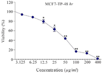
Figure 1. T. persicum extract reduces cancer cell viability. MCF-7 cells were treated with different concentrations of T. persicum extract for 48 hr, and then cell viability was measured by MTT test. The results are the mean±standard deviation of three separate experiments in which each treatment was repeated in 10 wells (*p<0.05; ** p<0.01; vs. control).
|
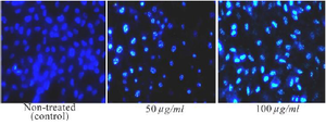
Figure 2. DAPI staining was performed on MCF-7 cells exposed to the ethyl acetate extract of T. persicum at concentrations of 50 and 100 μg/ml. MCF-7 cells treated with the extract showed apoptotic changes (condensed and fragmented nuclei). No such changes were observed in non-treated (control) cells (Magnification×40).
|
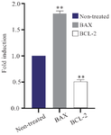
Figure 3. Real-time PCR experiments were conducted to measure the mRNA levels of the BAX and BCL2 genes in the MCF-7 cells, which were treated with the ethyl acetate extract of T. persicum. MCF-7 cells were treated with 50 μg/ml of the extract for 48 hr. Total RNA was collected from different sets of cells and was used for real-time PCR experiments. Data are normalized to the average mRNA levels of GAPDH. The shown data are mean±SD of three independent experiments (*p<0.05 and **p<0.01 vs. control).
|
|