Conjugation of Monoclonal Antibodies to Super Paramagnetic Iron Oxide Nanoparticles for Detection of her2/neu Antigen on Breast Cancer Cell Lines
-
Shamsipour, Fereshteh
-
Monoclonal Antibody Research Center, Avicenna Research Institute, ACECR , Tehran, Iran
-
Zarnani, Amir-Hassan
-
Department of Immunology, Reproductive Biotechnology Research Center, Avicenna Research Institute, ACECR , Tehran, Iran
-
Ghods, Roya
-
Monoclonal Antibody Research Center, Avicenna Research Institute, ACECR , Tehran, Iran
-
Chamankhah, Mahmood
-
Nanobiotechnology Research Center, Avicenna Research Institute, ACECR , Tehran, Iran
-
Forouzesh, Flora
-
Nanobiotechnology Research Center, Avicenna Research Institute, ACECR , Tehran, Iran
-
Vafaei, Sedigheh
-
Monoclonal Antibody Research Center, Avicenna Research Institute, ACECR , Tehran, Iran
-
Bayat, Ali Ahmad
-
Monoclonal Antibody Research Center, Avicenna Research Institute, ACECR , Tehran, Iran
-
Akhondi, Mohammad Mehdi
-
Department of Immunology, Reproductive Biotechnology Research Center, Avicenna Research Institute, ACECR , Tehran, Iran
-
Oghabian, Mohammad Ali
-
Research Centre for Science and Technology in Medicine, Tehran University of Medical Sciences , Tehran, Iran
-
 Jeddi-Tehrani, Mahmood
Ph.D., Monoclonal Antibody Research Center, Avicenna Research Institute, ACECR, Tehran, Iran, P.O. Box: 19615-1177, Tel: +98 21 22432020, Fax: +98 21 22432021, E-mail: mahjed@avicenna.ac.ir
Jeddi-Tehrani, Mahmood
Ph.D., Monoclonal Antibody Research Center, Avicenna Research Institute, ACECR, Tehran, Iran, P.O. Box: 19615-1177, Tel: +98 21 22432020, Fax: +98 21 22432021, E-mail: mahjed@avicenna.ac.ir
-
Monoclonal Antibody Research Center, Avicenna Research Institute, ACECR , Tehran, Iran
Abstract: Conjugation of monoclonal antibodies to super paramagnetic nanoparticles is an effective method for cancer diagnosis and treatment. In this study the humanized anti her2/neu monoclonal antibody- Herceptin- was conjugated to super paramagnetic iron oxide (SPIO) nanoparticles using EDC method. The concentration of the conjugated antibodies was measured by Bradford assay. The antibody-nanoparticle conjugates were incubated with SKBR-3 and T47D human breast carcinoma cell lines and the presence of the conjugates on cell surface was confirmed by Prussian blue iron staining method. Conjugation of Herceptin to SPIO resulted in a precipitate-free conjugate containing 20µg antibody/mg SPIO. Prussian blue iron-staining of cells showed successful binding of the conjugates to the cell surfaces. Conjugation of monoclonal antibodies to SPIO may be a useful method for detection of tumor cells, especially by MRI techniques.
Introduction :
Molecular probes for biomolecular recog-nition are of great importance in the fields of chemistry, biology, medical sciences and in biotechnology as well. These probes have been used in studies of biological functions and in ultrasensitive detection of biological factors responsible for many diseases (1). On the other front, the developments of nontoxic and biocompatible magnetic particles have been disclosed for biological applications since mid-1980s (2).
Recently, magnetic particles have attrac-ted growing interest as high performance biomaterial which is used for transport and separation of cells or cell parts (2, 3), MRI (4), hyperthermia (5) and drug delivery (6). Bio-logical samples such as blood, serum, cell suspensions and cell lysates are allowed to be exposed to specific ligand-coupled particles, and the captured molecules or cells are then rapidly separated using magnetic fields (3,7,8).
Magnetic particles conjugated with anti tumor monoclonal antibodies provide a new approach to identify tumor cells.
Antibodies labeled with magnetic nano-particles give magnetic signals on exposure to a magnetic field. Iron oxide particles are usually coated with different organic shells including dextran, albumin or polyethylene glycol. Coated nanoparticles can be manu-factured with a variety of functional groups (such as amino, aldehyde, hydroxyl, sulfate and carboxyl groups) on their surfaces. Con-sidering these properties, we used super para-magnetic iron oxide (nanomag-D-SPIO 20nm) with COOH group on the surface, for conjugation to a humanized anti her2/neu monoclonal antibody (Herceptin) as a cancer targeting antibody.
Materials and Methods :
Nanomag-D-SPIO 20 nm nanoparticles (surface COOH) and MACS separator with MS columns were purchased from Micromod (Miltenyi Biotech GmbH, Germany). The breast carcinoma cell lines SKBR-3 and T47D were obtained from Pasteur Institute of Iran. Other reagents and chemicals were obtained from Merck and Sigma.
Conjugation of anti her2 antibody (Herceptin) with nanoparticles by EDC method N-ethyl-N-(3-dimethyl aminopropyl) car-bodiimide hydrochloride (EDC, 26mM) and 10 mM N-hydroxy succinimide (NHS) were dissolved in 0.1 M 2-(N-morpholino) ethane- sulfonic acid (MES) buffer (pH=8.3). The mixture was added to 1 ml of 5 mg/ml nanomag-D-SPIO 20 nm nanoparticles, and shaken at room temperature for 2 hours.
The particles were washed twice with phosphate buffered saline (PBS) pH=7.4 and then 0.5-1 mg/ml of Herceptin was added to the activated particles. The mixture was sha-ken for 3 hours and the reaction was quenched by the addition of glycine for 30 minutes (9, 10).
The unconjugated antibodies were separ-ated from conjugated antibodies by MACS column. The amount of immobilized antibody was estimated based on the Bradford method.
Spectrophotometric measurement of Iron
Iron concentration of conjugated samples was obtained by potassium thiocyanate method (12,13). In brief, samples were diluted with 300 µl 6N HCl containing %1 H2O2; under this condition, the iron in the samples is dissolved and oxidized to ferric state. The samples were then added to a 5% solution of potassium thiocyanate where the Fe III formed a red complex with the thiocyanate which could be measured by absorbance at 480 nm.
Cell culture
The her2/neu expressing cell lines SKBR3 and T47D were grown in RPMI 1640 medium with 10% (v/v) fetal calf serum and %1 penicillin/streptomycin. Cells were incubated at 37°C containing 5% Co2.
Immunofluorescence staining
To verify the expression of her 2 proteins on the cells, the SKBR3 and T47D cells were incubated with anti her2/neu (Herceptin) at 10 µg/ml concentration for 1 hour at 37°C. After being washed in PBS, FITC-labeled anti human IgG (diluted 1/20, Avicenna Research Institute, Tehran, Iran) was added and incubated for 1 hour at room temperature. Cells were then observed directly on a fluo-rescence microscope (Olympus, Japan).
In vitro cell labeling
SKBR3 and T47D cells were counted and adjusted to a suspension of 4×10⁵cells/ml; 100 µl of each cell suspension were cyto-spined on microscope slides (Shandon cyto-spin 4, Thermo, Germany). The cells were incubated with 100 µl magnetic nanoparticles (with or without antibody; 5 µg Ab and 0.2 mg iron) for 1 hour at 37°C. Then cells were washed extensively with PBS to remove unbound particles. The nanoparticles that bounded on the cell surface were detected by iron staining with Prussian blue staining method (14, 15).
Result :
We used 20nm nanoparticles (a magnetic core covered with dextran) with carboxyl group for conjugation to Herceptin as a cancer targeting antibody. The final products of conjugation were suspensions without precipitate and the amount of immobilized antibody was 20-36 µg Ab/mg nanoparticles (Figure 1).
The amount of iron conjugated was deter-mined by potassium thiocyanate method using FeCl3-6H2O as a reference (Figure 2).
The SKBR3 and T47D cell lines showed high levels of her2/neu expression, after incubation with Herceptin-FITC (Figure 3), but not with the isotype control antibody (data not shown).
These results confirmed the presence of antigen on the cell surface. Specific binding of the conjugates to the cells can be deter-mined by using electron microscopy, MRI imaging and iron staining (13,18,19).
In this study after incubation of the cells with conjugated nanoparticles, the blue stain induced by iron staining showed the accu-mulation of conjugated nanoparticles on the cell surface (Figure 4).
Discussion :
Polymer-coated magnetic particles with particle sizes 5-500 nm have been employed in medicine or biotechnology for many years (16). In this study the 20 nm nanoparticles were coupled via their surface carboxyl group to the amino groups on the Herceptin antibody using the EDC method (10, 17).
After conjugation, the amount of immo-bilized antibody was approximately 20 µg/mg magnetite. However by increasing concentra-tion of antibody during the process, the efficiency of the conjugation did not improve. In other studies the efficiency of conjugation has been reported as 5-20 µg Ab/mg particles (9,18). Conjugated nanoparticles bound specific-ally to the her2/neu antigen.
Iron staining, confirmed the presence of nanoparticles on the cell surface. In the present study we showed specific binding of Herceptin-nanomagnetic particle conjugates to her2/neu over expressing cells, suggesting a future application of Herceptin-magnetite for MR imaging of breast cancer.
Acknowledgement :
This work was supported by a grant from the Nanotechnology Committee of Iran’s Ministry of Health and Medical Education.

Figure 1. Antibody concentration measurement by Bradford assay (Ab conc. 100 �g/ml)
|
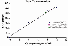
Figure 2. Magnetic nanoparticles concentration measurement by Potassium Thiocyanate method (particles conc. 4-4.5mg/ml)
|
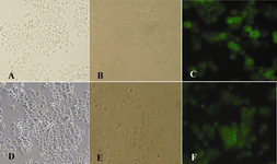
Figure 3. Light microscopy and Immunoflurescence of SKBR3 and T47D cell lines A: SKBR3 cells in culture, B: suspension of SKBR3 cells, C: SKBR3 cells after incubation with Herceptin-FITC, D: T47D cells in culture, E: suspension of T47D cells, F: T47D cells after incubation with Herceptin- FITC.
|
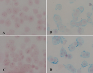
Figure 4. Iron staining of cells after incubation with conjugated nanoparticles A: SKBR3 cells, B: KBR3 with iron staining, C: T47D cells, D: with iron staining.
|
|