Toxicity Study of Silver Nanoparticles Synthesized from Suaeda monoica on Hep-2 Cell Line
-
Satyavani, Kaliyamurthi
-
Marine Medicinal Plant Biotechnology Laboratory, Faculty of Marine Sciences, Annamalai University, Parangipettai, Tamil Nadu, India
-
Gurudeeban, Selvaraj
-
Marine Medicinal Plant Biotechnology Laboratory, Faculty of Marine Sciences, Annamalai University, Parangipettai, Tamil Nadu, India
-
 Ramanathan, Thiruganasambandam
Thiruganasambandam Ramanathan, Ph.D., Marine Medicinal Plant Biotechnology Laboratory, Faculty of Marine Sciences, Annamalai University, Parangipettai, Tamil Nadu, India, E-mail: marinepharmacology@gmail.com
Ramanathan, Thiruganasambandam
Thiruganasambandam Ramanathan, Ph.D., Marine Medicinal Plant Biotechnology Laboratory, Faculty of Marine Sciences, Annamalai University, Parangipettai, Tamil Nadu, India, E-mail: marinepharmacology@gmail.com
-
Marine Medicinal Plant Biotechnology Laboratory, Faculty of Marine Sciences, Annamalai University, Parangipettai, Tamil Nadu, India
-
Balasubramanian, Thangavel
-
Marine Medicinal Plant Biotechnology Laboratory, Faculty of Marine Sciences, Annamalai University, Parangipettai, Tamil Nadu, India
Abstract: Recently there has been fabulous excitement in the nano-biotechnological area for the study of nanoparticles synthesis using some natural biological system, which has led the growth advanced nanomaterials. This intention made us to assess the biologically synthesized silver nanoparticles from the leaf of Suaeda monoica (S.monoica) using 1 mM silver nitrate. The leaf extract of S.monoica incubated with 1 mM silver nitrate solution and characterized by UV- spectrometer and AFM. The effect of synthesized silver nanoparticles on Human Epidermoid Larynx Carcinoma cell line was evaluated by the MTT colorimetric technique. As a result we observed gradual change in the colour of extract from greenish to brown. The synthesized silver nanoparticles con-firmed by UV at 430 nm and spherical shape identified in the range of 31 nm under AFM. The effect of silver nanoparticles on Human Epidermoid Larynx Carcinoma cell line exhibits a dose-dependent toxicity for the cell tested and the viability of Hep-2 cells decreased to 50% (IC50) at the concentration of 500 nM. Further findings will be determined the exact mechanisms of this cost effective Nano-treatments.
Introduction :
Suaeda monoica is a salt marsh herb grow-ing in hyper saline soils that belongs to Chen-opodiaceae and is distributed throughout the East and West coast mangroves in India. Traditionally, the leaf from S.monoica is known to be used as a medicine for hepatitis and scientifically it is reported to be used as ointment for wounds and possess antiviral activity because of the presence of triterpe-noids, sterols (1).
Rapidly developing field of nanoscience had raised the possibility of using therapeutic nanoparticles in the diagnosis and treatment of human cancers (2). Nanoscale particles and molecules are a potential alternative for treat-ment of disease because they have unique biological effects based on the structure and size, which differ from traditional small molecule drugs (3).
In the last few years, several pharmaceut-icals companies had obtained approval from the US Food and Drug Administration (FDA) for the development of nanotechnology based drugs. Silver had long been recognized as having an inhibitory effect towards many bac-terial strains and micro organisms commonly present in medical and industrial processes (4). The most widely used and well known appli-cations of silver and silver nanoparticles in-cludes topical ointments and creams contain-ing silver to prevent infection of burns and open wounds (5).
Production of nanoparticles can be achieved through different methods. Chemical approaches are the most popular methods for the production of nanoparticles. However, some chemical methods cannot avoid the use of toxic chemicals in the synthesis protocol. Since noble metal nanoparticles such as gold, silver and platinum nanoparticles are widely applied to human contacting areas, there is a growing need to develop environmentally friendly processes of nanoparticles synthesis that do not use toxic chemicals. Biological methods of nanoparticles synthesis using microorganisms (6), enzyme (7), and plant or plant extract have been suggested as possible ecofriendly alternatives to chemical and phys-ical methods. Using plant for nanoparticles synthesis can be advantageous over other bio-logical processes by eliminating the elaborate process of maintaining cell culture (8).
The role of silver nanoparticles as an anti-cancer agent should open new door in the field of medicine. Silver nanoparticles should serve as one of the best ways of treating dise-ases that involve cell proliferation and cell death (9). In the present study, we screened salt marsh plant species S.monoica leaf extracts for extracellular silver nanoparticles synthesis and to determine the cytotoxicity threshold in this Human Epidermoid Larynx Carcinoma (Hep-2) cell line.
Materials and Methods :
Plant material
S.monoica leaves freshly were collected from the Kollidam coast (Tamil Nadu) India. The specimen was certified by Botanical Sur-vey of India (BSI) Coimbatore, and docu-mented in the Herbaria of C.A.S. in Marine Biology (Voucher No. AUCASMB15), Anna-malai University, India.
Synthesis of silver nanoparticles
The analytical grade silver nitrate was pur-chased from Sigma Chemicals (Mumbai). The leaves weighing 25 g were thoroughly washed in distilled water, dried, cut into fine pieces and were crushed into 100 ml sterile distilled water and filtered through what man No.1 filter paper. The filtrate was further filtered through 0.6 μm sized filters and stored at 4 ºC. Ten ml suspension of leaf extract was added to 90 ml aqueous solution of silver nitrate
(1 mM) solution separately for reduction in to Ag+ ions and incubated at room temperature (35 °C) for 5 hr. After 5 hr of incubation the silver nanoparticles were isolated and concen-trated by repeated (4-5 times) centrifugation of the reaction mixture at 10,000×g for
10 min. The supernatant was replaced by dis-tilled each time and suspension stored as lyo-philized powder. The optical measurements, was carried out by UV-Vis spectrophotometer (UV- 2450 (Shimadzu) and scaned the spectra between 200-700 nm at the resolution of 1nm.
Atomic force microscope
Purified SNP (Silver Nanoparticles) in sus-pension also characterized their morphology using a VEeco diNanoscope 3D AFM (Atomic Force Microscope). A small volume of sam-ple was spread on a well-cleaned glass cover slip surface mounted on the AFM stub, and was dried with nitrogen flow at room tem-perature.
Images were obtained in tapping mode using a silicon probe cantilever of 125 μm length, resonance frequency 209-286 kHz, spring constant 20-80 nm-1 minimum of five images for each sample were obtained with AFM and analyzed to ensure reproducible results.
Cell culture
The Hep-2 cell line was purchased from National Cell Centre, Pune (India). Cancerous cells were seeded in flask with MEM (Mini-mal Essential media) medium with 2-10% Fetal Calf Serum (FCS) and incubated at 37°C in a 5% CO2. After 24 hr incubation period the attached cells were trypsinizated for 3-5 min and centrifuged (1,400 rpm,
5 min). The cells were counted and distributed in 96 well ELISA plate with 10,000 cells in each well. The plate was incubated 24 hr at 37 C in a 5% CO2 atmosphere to allow the cells attach to the bottom of the well (10).
Cell treatment with silver nanoparticles
Silver nanoparticles reduced ATP content of the cell, caused damage to mitochondria and increased production of Reactive Oxygen Species (ROS) in a dose-dependent manner (10). Hence we determined the toxicity of silver nanoparticles at different concentrations (15, 30, 62, 125, 250, 500, 1000 µg/ml) and dupli-cates added to the wells were incubated in to as grown cell (1×10⁴ cells/well) and the cell population was determined by optical micro-scopy at 24 and 48 hr.
MTT assay
Cell viability was evaluated by the MTT colorimetric technique with slight modifica-tion (11). Briefly, in each well 200 µl of MTT [3-(4, 5-dimetheylthiazol-2)-2, 5 diphenyl tetrazolium bromide] without phenol red, yel-lowish in color solution (5 mg/ml in PBS), was added to each well. The plates were in-cubated for 6-7 hr in 5% CO2 incubator for reduction of MTT by metabolically active cells, in part by the action of dehydrogenase enzymes, to generate reducing equivalents such as NADH and NADPH. The resulting intracellular purple formazon solubilized the MTT crystals by adding and quantified by spectrophotometric mean and then the super-natants were removed.
For solubilization of the MTT crystals,
100 µl of isoproponal or DMSO was added to the wells. The plates were placed on a shaker for 15 min for complete solubilization of crys-tals and then the optical density of each well was determined. The quantity of formazan product, as measured by the amount of
595 nm using a scanning Multiwell spectro-photometer (Biorad, Model 680, Japan) and DMSO, served as a blank. The absorbance is directly proportional to the number of living cells in cultural.
Each experiment was done in duplicate. The relative cell viability (%) related to con-trol wells containing cell culture medium without nanoparticles as a vechicle was cal-culated by (A) test/ (A) control×100; where (A) test is the absorbance of the test sample and (A) control is the absorbance of control sample.
Statistical analysis
All experiments were done in duplicate, and the results were presented as mean± standard deviation. The experimental data were analyzed by using SPSS. Statistical sig-nificance was accepted at a level of p<0.05 to calculate IC50 values.
Results :
The leaf extract of S.monoica was used for the synthesis of silver nanoparticles. The re-action started within first hr of the incubation with silver nitrate (1 mM). The primary detec-tion of synthesized silver nanoparticles was carried out in the reaction mixture by ob-serving the colour change of the medium from greenish to dark brown. The reaction rate was maximum after 25 hr of incubation as in-dicated by the formation of silver nanopar-ticles.
Our findings showed resemblance to the re-sults already reported by in the case of extract of Capsicum annum (12) and in case of extract of Aloe vera (13). They reported that when the extracts of their respective test plants were challenged with silver nitrate (1 mM), it turn-ed brown and the intensity of colour was in-creased with the time of incubation.
In order to verify the synthesis of silver nanopartices, the test samples were subjected to the UV-Vis spectrophotometer analysis after 5 hr of incubation. A peak specific for the synthesis of silver nanoparticles was ob-tained at 430 nm. AFM is used for morpho-logical characterization of SNP (14). The shape of the SNP synthesized by leaf extract was spherical and was found to be in the range of 31 nm (Figure 1).
The in vitro cytotoxicity effects of silver nanoparticles were screened against human epidermoid larynx carcinoma cell lines by means of MTT assay. The silver nanoparticles were able to reduce viability of the Hep-2 cells in a dose-dependent manner, as shown in
figure 2. After five hr of treatment, the silver nanoparticles were found to be cytotoxic to tumour cells at a concentration of 500 nM and higher. Silver nanoparticles at 500 nM de-creased the viability of Hep-2 cells (Figure 3) to 50 % of the initial level, and this was chosen as the IC50. Longer exposures resulted in additional toxicity to the cells.
These results demonstrate that silver nano-particles mediate a dose and time dependent increase in toxicity. The result suggested that the plant mediated synthesized silver nano-particles possess great selectivity to cancer cell and can display potential application in cancer chemoprevention and chemotherapy. Silver nanoparticles revealed to have import-ant anti angiogenic properties (15); so are at-tractive for study of their potential antitumor effects. Compounds possessing antiangio-genic properties are known for their potential ability to block the activity of abnormally ex-pressed signaling proteins (16).
Discussion :
In the present study, it has been shown a simple, rapid and green synthesize of silver nanoparticles from S.monoica of size 31 nm, at a concentration of 500 nM had cytotoxic effects on Hep-2 tumor cells under in vitro conditions. Similar reports was observed by Bilberg et al, in male zebra fish with a semi-static 48 hr exposure LC50 of 84 μg L−1 and LC10 of 57 μg L−1 (17). Initially, a dose de-pendent effect of SNP on Hep-2 cell lines assessed by MTT assay showed an IC50 value of about 500 nM that induced partial reduc-tion in cell viability in comparison with control.
The cytotoxic effect of SNP on cell viabil-ity has a major role in antitumor activity, thereby reducing disease progression. The cy-totoxic effects of silver are the result of active physiochemical interaction of silver atoms with functional groups of intracellular pro-teins, as well as with the nitrogen bases and phosphate groups in DNA (18).
Conclusion :
In conclusion, the silver nanoparticles syn-thesized from S.monoica serve as an anti-tumor agent by decreasing progressive devel-opment of tumor cells. Our result suggests that SNP can induce cytotoxic effects on Hep-2 cells, inhibiting tumour progression and thereby effectively controlling progression without toxicity to normal cells. This may be due to their inhibitory activities in several sig-naling cascade responsible for the develop-ment and pathogenesis of disease which are as yet not understood.
Although, some of the researchers pointed out that the nanosilver toxicity is caused by chemical interactions, the toxic portion of nanoparticles must originate either from silver ions dissolved from the particle or from the exposed silver atoms on the particle surface (19). While the mechanism(s) by which AgNPs are toxic are unclear, their increasing use raises the concern that its release into the environment could lead to environmental toxicity.
Acknowledgement :
The authors are gratefully acknowledge to the Dean, Faculty of Marine Sciences, Anna-malai University, Parangipettai, Tamil Nadu, India for providing all support during the study period. The authors gratefully acknow-ledge the Dean, Faculty of Marine Sciences, Annamalai University, Parangipettai, Tamil Nadu and DST-PURSE program, India for financial support during the study period.
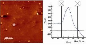
Figure 1. Representative AFM image of nanoparticles synthesized from different plant species (Citrullus colocynthis and Suaeda monoica) using callus and leaf extracts. Despite originating from distinct plant sources, the nanoparticles exhibit uniform morphology and an average size of ~31 nm. The consistency in nanoparticle size and shape supports the reproducibility of the synthesis process.
|
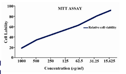
Figure 2. Cytotoxic study of silver nanoparticles synthe-sized from leaf of Suaeda monoica against Hep-2 cell line on a dose dependent manner
|
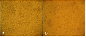
Figure 3. Cytotoxicity of Silver Nanoparticles (SN) syn-thesized from leaf of leaf of Suaeda monoica on Hep2 cell line (A) Normal cells (B) SN treated cells (500 �g/ml) de-creased the viability of HeP-2 cells
|
|