Identification of a Novel Nucleic Acid Target for the Rapid and Specific Detection of Mycobacterium Simiae Using Comparative Genomic Analysis
-
Abavisani , Mohammad
-
Student Research Committee, Mashhad University of Medical Sciences, Mashhad, Iran
-
Ghazvini , Kiarash
-
Department of Microbiology and Virology, Faculty of Medicine, Mashhad University of Medical Sciences, Mashhad, Iran
-
Zare, Hosna
-
Student Research Committee, Mashhad University of Medical Sciences, Mashhad, Iran
-
Department of Laboratory Sciences, School of Paramedical Sciences, Mashhad University of Medical Sciences, Mashhad, Iran
-
Hojatpanah , Noshin
-
Student Research Committee, Mashhad University of Medical Sciences, Mashhad, Iran
-
Department of Microbiology and Virology, Faculty of Medicine, Mashhad University of Medical Sciences, Mashhad, Iran
-
Falahi , Jamal
-
Department of Laboratory Sciences, School of Paramedical Sciences, Torbat Heydariyeh University of Medical Sciences, Torbat Heydariyeh, Iran
-
 Neshani, Alireza
Department of Laboratory Sciences, School of Paramedical Sciences, Mashhad University of Medical Sciences, Mashhad, Iran, Tel: +98 9395100919, 513 8829278; E-mail: Neshania@mums.ac.ir, alireza.neshani@gmail.com
Neshani, Alireza
Department of Laboratory Sciences, School of Paramedical Sciences, Mashhad University of Medical Sciences, Mashhad, Iran, Tel: +98 9395100919, 513 8829278; E-mail: Neshania@mums.ac.ir, alireza.neshani@gmail.com
-
Student Research Committee, Mashhad University of Medical Sciences, Mashhad, Iran
Abstract: Background: Mycobacterium simiae (M. simiae) is a non-tuberculous mycobacterium (NTM) that closely resembles Mycobacterium tuberculosis (M. tuberculosis) in clinical and biochemical characteristics, notably its niacin-positive phenotype. This similarity frequently leads to misdiagnosis and inappropriate treatment with first-line anti-tuberculosis drugs, to which M. simiae is frequently resistant. Current diagnostic methods are expensive or need complex equipment, highlighting the urgent need for a rapid, specific, and accessible molecular target to identify M. simiae accurately.
Methods: In this study, a modified genome comparison method was applied to the complete reference genome of M. simiae (AP022568.1) in order to identify a putative species-specific nucleotide sequence. A conventional PCR assay was designed to amplify a 168-bp fragment within this target, designated MST601 (M. simiae Target, 601 bp). The analytical sensitivity [Limit of Detection (LOD)] was determined using serial dilutions of genomic DNA. The pilot evaluation of the assay was assessed using 10 well-characterized clinical isolates of M. simiae.
Results: The MST601-PCR assay demonstrated high analytical sensitivity, with a limit of detection of ~10 fg (≈2 genome equivalents per reaction) of M. simiae genomic DNA. No cross-reactivity among the tested species was observed with any of the 10 non-target mycobacterial species tested. The assay successfully amplified the target sequence from all 10 clinical isolates. Sequencing of the amplicons revealed ≥99% identity to reference M. simiae strains in the GenBank database, validating the assay's accuracy.
Conclusion: A species-specific nucleic acid target, MST601, facilitating the rapid and accurate detection of M. simiae via conventional PCR was presented. This assay pro-vides a low-cost and accessible option for diagnostic laboratories.
Introduction :
Non-Tuberculous Mycobacteria (NTM) constitute a notable category of environmental micro-organisms that have garnered heightened interest in clinical microbiology owing to their potential to induce opportunistic infections, especially in immunocompromised patients and individuals with pre-existing pulmonary disorders 1. Within the over 200 species classified in the NTM group, Mycobacterium simiae (M. simiae) is distinguished as a distinct pathogen due to its slow growth, non-pigmented colonies, and particularly its niacin-positive feature, a property it shares exclusively with Mycobacterium tuberculosis (M. tuberculosis; MTB). The biochemical resemblance frequently results in diagnostic ambiguity between M. simiae and MTB, complicating clinical management and therapy choices 2.
M. simiae is an emerging pathogen predominantly linked to pulmonary infections, particularly in individuals with pre-existing lung conditions such as Chronic Obstructive Pulmonary Disease (COPD), bronchiectasis, or cystic fibrosis 3,4. Historically perceived as geographically confined to areas like Cuba, Gaza, and the southern United States, current reports reveal its occurrence in several more places worldwide, including the Middle East 2,5. A meta-analysis study indicated that between 1993 and 2012, the prevalence of NTM infections among Tuberculosis (TB) suspects in Iran was approximately 10.2%, with 43.3% of these cases attributed to M. simiae 6. During the same period, the World Health Organization (WHO) reported an incidence rate of TB in Iran ranging from 18 to 54 cases per 100,000 population 7,8. The clinical signs of M. simiae infections closely resemble those of TB, rendering precise diagnosis essential yet challenging 9.
Accurate identification of M. simiae is further complicated by the potential for misdiagnosis with other NTM, particularly those sharing similar growth characteristics or biochemical profiles. For instance, M. simiae may be misidentified as members of the Mycobacterium avium Complex (MAC) or other slow-growing NTM due to overlapping clinical presentations and radiological features 10. This risk is especially critical in regions where multiple NTM species coexist and in cases of co-infection, where the presence of more than one mycobacterial species can lead to diagnostic ambiguity and suboptimal treatment strategies. Failure to distinguish M. simiae from other NTM in such scenarios may result in the inappropriate use of macrolide-based regimens, which are ineffective against M. simiae due to its frequent resistance 11,12. Therefore, a highly specific diagnostic tool is essential not only to differentiate M. simiae from M. tuberculosis but also to accurately identify it in the context of mixed or co-occurring NTM infections. The absence of a widely accepted, species-specific genomic target for M. simiae suitable for simple PCR-based detection has limited the development of efficient and uncomplicated molecular diagnostic methods like Polymerase Chain Reaction (PCR) or Loop-mediated isothermal amplification (LAMP) 5.
Conventional diagnostic approaches reliant on culture techniques and phenotypic attributes, such as colony morphology, growth rate, pigment synthesis, and biochemical assays, are not only laborious, but also challenging to interpret and frequently unreliable 13. Despite the development of various advanced molecular techniques for the detection of M. simiae, including Restriction Fragment Length Polymorphism (RFLP) 14, real-time PCR 15, Matrix-Assisted Laser Desorption/Ionization Time-of-Flight Mass Spectrometry (MALDI-TOF MS) 16, line probe assays 13,17, sequencing 15, and electrochemical label-free DNA Nanobiosensors 5, these methods are considerably more costly than traditional PCR-based assays and necessitate highly skilled personnel. Importantly, most of these assays either target conserved loci (e.g., 16S rRNA, hsp65, rpoB) that do not provide unequivocal discrimination of M. simiae from closely related NTM, or depend on specialized, high-cost platforms that are not universally accessible. Although conventional PCR is a well-established and routine technique in molecular microbiology, its widespread availability, low cost, and modest equipment requirements make it particularly suitable for laboratories in low-and middle-income countries and regional TB reference centers. Therefore, identifying a robust, species-specific genomic target that can be detected using conventional PCR has substantial practical value for the accurate diagnosis of M. simiae.
In light of these challenges, there is an urgent need for cost-effective, accessible, and highly precise diagnostic tools to distinguish M. simiae from other mycobacterial infections. The lack of a specific nucleic acid diagnostic target for M. simiae intensifies this problem, necessitating the development of novel markers suitable for straightforward, cost-effective PCR-based assays. To address this gap, a comparative genomic analysis, a method previously established by Neshani et al 18,19 was applied, to identify a nucleotide sequence that appears species-specific to M. simiae based on comparative genomic analysis. The primary novelty of this study lies in the identification and validation of this new target and its translation into a simple assay that can be implemented using conventional PCR, a platform that is already routinely available in most clinical and reference laboratories. The specific objectives of this study were: (1) to identify a candidate genomic region through in silico genome comparison; (2) to design and optimize a conventional PCR assay for the detection of this target; and (3) to evaluate the analytical sensitivity, analytical specificity, and performance of the PCR assay on reference strains and well-characterized clinical isolates.
Materials and Methods :
This study utilized comparative genomic analysis to discover a species-specific genomic target in M. simiae, which has a genome size of around 5.7 million base pairs 18. To reduce analytical errors and guarantee precision, the complete target-identification workflow was allocated among four researchers, with each step executed independently and cross-validated. This organization ensured that the genome-wide comparative analysis was performed in a systematic and reproducible manner, rather than as an ad hoc or single BLAST search.
Identifying specific target: The entire genome sequences of M. simiae were compared with the genomic data accessible in the NCBI database to ascertain the most precise and dependable target sequence. The workflow was executed as follows: Initially, all accessible M. simiae genomic sequences in the NCBI database were evaluated, and a reference quality sequence, ideally from the RefSeq database, was chosen as a starting point for subsequent research (Table 1). The M. simiae genome with accession number AP022568.1 was used as the reference sequence for this work.
This choice was predicated on its categorization within the NCBI RefSeq database, which offers fully assembled, high-quality genomes accompanied by standardized and meticulously reviewed annotations by the NCBI database. In comparison to other sequences classified as WGS (Whole Genome Shotgun) or GenBank entries—some of which may be incomplete or of inferior quality—the AP022568.1 sequence provides enhanced reliability and precision. Consequently, it was considered the most appropriate genomic reference for executing accurate comparative analyses of M. simiae.
The genomic sequence of M. simiae was obtained from the NCBI database and divided into about 5000 bp segments, yielding a total of 1157 pieces. Each fragment underwent independent analysis using BLASTn against the NCBI Nucleotide collection (nt) via the online BLASTn interface (https://blast.ncbi. nlm.nih.gov/Blast.cgi) between March and May 2024. The BLAST findings were thoroughly analyzed to identify the most distinctive segments unique to M. simiae. Two primary factors were utilized in this selection process: all four selected M. simiae strains enumerated in table 1 must appear in the BLAST results with both a sequence identity and query coverage of at least 90%. No additional bacterial species should exhibit a query coverage of over 10% in the BLAST output. As BLAST results are inherently organized by query coverage in descending order, non-target organisms exhibiting greater resemblance can be readily discerned towards the conclusion of the list. The most specific genomic area was chosen based on these studies. A subsegment of this region exhibiting 100% specificity was selected and designated as MST601 (Mycobacterium simiae Target, 601 bp) (Figure 1). This approach represents a systematic, genome-wide screening rather than the selection of a single candidate gene. By fragmenting the complete reference genome into 1157 overlapping segments and testing each fragment against the entire NCBI nucleotide collection, the aim was to minimize bias with focusing only on commonly used loci (e.g. 16S rRNA, hsp65, rpoB). The stringent dual thresholds (≥90% identity and coverage for all available M. simiae strains and ≤10% query coverage for any non-M. simiae species) were chosen to reduce the risk of cross-reactivity even in the presence of incomplete or misannotated genomes. This genome-wide, threshold-based strategy allowed the unbiased identification of MST601 as a highly specific marker for M. simiae.
In silico specificity study of the MST601 sequence: An in silico alignment study was conducted to assess the species specificity of the MST601 sequence. The whole nucleotide sequence of MST601 was individually compared with the genomes of phylogenetically related mycobacterial species. Reference genome sequences were acquired from the NCBI database. The MST601 sequence for each selected species was aligned using the "NCBI BLAST 2 Sequences (BLASTn)" tool. Accessible online at: (https://blast. ncbi.nlm.nih.gov/Blast.cgi?PROGRAM=blastn&PAGE_TYPE=BlastSearch&USE_DEFAULTS=on&BLAST_SPEC=blast2seq). Alignment parameters including query coverage, percentage identity, and match count were documented. This method facilitated a focused evaluation of probable homology between MST601 and non-target Mycobacterium genomes.
The selected panel of species included both phylogenetically related mycobacteria and clinically relevant non-tuberculous mycobacteria that are commonly encountered in pulmonary infections. The reference genomes and their accession numbers are listed in table 2. This second-level, targeted in silico validation complements the initial genome-wide screening and provides an additional layer of assurance that MST601 does not share substantial homology with non-target Mycobacterium species.
Primer design and PCR protocol: The design and analysis of primers targeting the M. simiae specific genomic region MST601 were conducted using Oligo7 and the OligoAnalyzer™ Tool version 3.1 (https://eu.idtdna.com/calc/analyzer) (Integrated DNA Technologies, Coralville, IA, USA). The primer combination is engineered to amplify a 168 bp fragment, ensuring enhanced specificity and efficiency in PCR detection. The primer sequences and amplicon information are presented in figure 1.
The PCR was conducted in a total volume of 25 μl, comprising 1 μl of sample DNA, 2.5 μl of 10×PCR buffer (100 mM Tris-HCl [pH=8.3], 500 mM KCl), 1.5 μl of 25 mM MgCl2, 0.5 μl of 200 μM of each of the four dNTPs, 1 μl of each 10 μM forward and reverse primers, PCR-grade water, and 0.625 U of Taq DNA polymerase. The DNA amplification was conducted using a Thermal Cycler (Techne TC-3000X, Stone, Staffordshire, UK). The procedure commenced with an initial denaturation at 95°C for 5 min, followed by 40 cycles consisting of (i) denaturation at 95°C for 15 s, (ii) annealing at 61°C for 20 s, (iii) extension at 72°C for 30 s, and concluded with a final extension at 72°C for 5 min.
Then, 5 μl of PCR product was electrophoresed on a 1.5% agarose gel prepared in 1×TAE buffer and containing DNA Green Viewer (Parstous, Tehran, Iran). Gels were run at 90 V for 45 min using an Owl EasyCast B1 electrophoresis system (Thermo Fisher Scientific, Waltham, MA, USA), alongside a GeneRuler 100 bp DNA Ladder (Thermo Fisher Scientific) as a size marker. DNA bands were visualized under ultraviolet illumination using a Spectroline UV transilluminator (Spectroline, New York, NY, USA). The detection of a 168 bp fragment signified a favorable outcome. Ten percent of positive specimens were randomly selected, and sequencing was conducted on their amplicons. To ensure assay reliability, each PCR run included a No-Template Control (NTC) with nuclease-free water instead of DNA template, and an extraction blank processed alongside samples during DNA isolation. Both controls consistently yielded negative results, confirming the absence of contamination. Given that all tested samples were purified genomic DNA from cultured isolates (not crude clinical specimens), the risk of PCR inhibition was minimal, and thus an Internal Amplification Control (IAC) was not included in this validation study.
Analytical sensitivity (detection limit): Genomic DNA of M. simiae (verified isolate, stored in the microbial culture collection of the Department of Laboratory Sciences, Mashhad University of Medical Sciences) was extracted from cultured colonies using the commercial mycobacterial DNA extraction kit (Karmania Pars Gene, Iran; Cat. No. KPG-DNAMTB), which is based on silica-membrane technology for efficient recovery of mycobacterial DNA. The protocol was performed according to the manufacturer’s instructions. The concentration of DNA was measured utilizing a NanoDrop ND-1000 Spectrophotometer (NanoDrop, Fisher Thermo, Wilmington, DE, USA). Serial dilutions of genomic DNA were created in nuclease-free distilled water to obtain concentrations of 100 pg, 10 pg, 1 pg, 100 fg, 10 fg, 5 fg, and 1 fg/μl, with 1 μl of each dilution utilized as a template in PCR experiments. The sensitivity of MST601-PCR was assessed by identifying the minimum concentration that produced a visible amplicon on agarose gel electrophoresis. Each dilution was evaluated on three distinct days to determine repeatability. Assuming a genome size of approximately 5.7 Mb for M. simiae 18 and an average molecular weight of 660 Da per base pair of double-stranded DNA, 10 fg of genomic DNA per reaction corresponds to roughly two genome equivalents.
Analytical specificity: To assess the analytical specificity of MST601-PCR, genomic DNA from M. simiae and other closely related mycobacterial species, including M. tuberculosis, Mycobacterium bovis (M. bovis) BCG, Mycobacterium smegmatis (M. smegmatis), Mycobacterium kansasii (M. kansasii), Mycobacterium avium (M. avium), Mycobacterium chelonae (M. chelonae), Mycobacterium fortuitum (M. fortuitum), Mycobacterium ulcerans (M. ulcerans), Mycobacterium intracellulare (M. intracellulare), and Mycobacterium marinum (M. marinum) was utilized as templates in PCR assays (verified isolate, preserved in the microbial culture collection of the Department of Laboratory Sciences, Mashhad University of Medical Sciences). A quantity of 1 ng of genomic DNA per reaction was used for all tested species to ensure standardized conditions during specificity evaluation.
The PCRs were conducted under the same circumstances as those employed for sensitivity testing. Specificity was deemed adequate when amplification occurred solely in samples containing M. simiae DNA, with no amplification observed for any of the non-target mycobacterial species tested.
Assay performance on DNA from clinical isolates: Genomic DNA was taken from a collection of 10 verified clinical isolates of M. simiae, which had been obtained from sputum samples of patients suspected for TB, and previously cultured and confirmed via ITS sequencing. The isolates were stored in the microbial culture collection of the Department of Laboratory Sciences at Mashhad University of Medical Sciences. All DNA samples performed MST601-PCR following the previously outlined methodology. Amplification products were examined by agarose gel electrophoresis under the same conditions described in section 2.3.
To test the specificity of MST601-PCR, amplicons from all ten M. simiae clinical isolates were sequenced utilizing forward and reverse primers. Sequencing reactions were conducted using an automated DNA sequencer. Amplicon sequencing was performed by the BGI Group (Beijing Genomics Institute, Shenzhen, China). The produced sequences were examined with the BLASTN program (https://blast.ncbi.nlm.nih.gov/) for comparison to the reference sequences in the GenBank database.
Results :
Target finding: The 601 bp fragment encompassing nucleotides 4,431,077 to 4,431,677 of M. simiae JCM 12377 (AP022568.1) was recognized as the most specific sequence (Figure 1) (GenBank: accession number: PV878584). The BLASTN search indicated that within the NCBI registered complete genomes (nucleotide collection database), this sequence could specifically identify all strains of M. simiae.
In silico specificity analysis of the MST601 sequence: The alignment analysis revealed that the MST601 sequence was solely found in M. simiae and exhibited no substantial similarity to the genomes of other mycobacterial species analyzed (Table 2). No comprehensive or highly conserved matches were identified among the non-target species, thereby affirming the specificity of MST601 for M. simiae. A limited number of non-simiae species exhibited partial alignments; however, these correspondences constituted less than 5% of the overall sequence length and demonstrated identities below 93%, signifying negligible cross-reactivity.
Analytical sensitivity (detection limit): The analytical sensitivity of MST601-PCR was assessed using serial dilutions of M. simiae genomic DNA. Clear and consistent amplification was observed for DNA concentrations ranging from 100 pg to 10 fg/μl, as indicated by the presence of a distinct band of the expected size on agarose gel electrophoresis (Figure 2). At the lowest concentrations tested (5 fg and 1 fg), no visible amplicon was detected under UV transillumination, establishing the detection limit of the assay at approximately 10 fg of genomic DNA per reaction (equivalent to approximately 1.5 copies of M. simiae genomic DNA per μl). All dilutions were tested in triplicate across three independent runs, demonstrating the high reproducibility of the assay on different experimental days.
Analytical specificity: The analytical specificity of MST601-PCR was evaluated using genomic DNA from M. simiae and closely related mycobacterial species, including M. tuberculosis, M. bovis BCG, M. smegmatis, M. kansasii, M. avium, M. chelonae, M. fortuitum, M. ulcerans, M. intracellulare, and M. marinum. Amplification occurred solely in samples containing M. simiae DNA, yielding a distinct 168 bp band on agarose gel electrophoresis (Figure 3). No amplicon was identified in any of the non-simiae mycobacterial species tested, demonstrating the high specificity of the MST601-PCR assay for M. simiae.
Assay performance on DNA from clinical isolates: In a pilot clinical evaluation, MST601-PCR effectively amplified the target sequence from all ten verified clinical isolates of M. simiae. Electrophoretic analysis demonstrated a specific 168 bp amplicon in every sample, with no amplification seen in negative controls (Figure 4). The results validate the assay’s excellent sensitivity in detecting M. simiae DNA among a range of clinical isolates. Analysis of all 10 amplicons demonstrated ≥99% identity with M. simiae reference strains in GenBank, hence validating the specificity of the MST601-PCR test. It is important to note that this assessment evaluates analytical performance on purified DNA from isolates and should not be interpreted as clinical sensitivity, which would require testing of direct patient specimens.
Discussion :
The precise and prompt identification of M. simiae is essential for optimal clinical care, especially because of its phenotypic and biochemical resemblances to M. tuberculosis 20. This study identifies MST601 as the nucleotide sequence identified for M. simiae, enabling the rapid and specific detection of this pathogen via a conventional PCR assay. This strategy resolves a significant diagnostic deficiency in contemporary laboratory operations, where protracted culture techniques and expensive molecular assays frequently postpone diagnosis and complicate treatment choices. In this study, aim was not to introduce a new amplification technology. Instead, the main contribution was the identification of MST601 as a highly specific genomic marker for M. simiae and its implementation in a conventional PCR format. While conventional PCR is a routine method in molecular microbiology, its broad availability, minimal equipment demands, and low cost make it a practical backbone for diagnostics in many clinical laboratories, particularly in resource-limited settings. By coupling this widely accessible platform with a newly defined species-specific target, the present work addresses a critical gap in the molecular diagnosis of M. simiae.
Although M. avium and M. intracellulare are the most common NTM species globally, M. simiae demonstrates notable regional prevalence 21,22. In Iran, it accounts for up to 43% of NTM isolates, emphasizing the need for region-specific diagnostic tools 6. The MST601-PCR assay is particularly valuable in such endemic areas where access to advanced molecular diagnostics is limited.
Genome comparison methods have identified highly specific genetic sequences for molecular diagnostics, improving the sensitivity and specificity of PCR-based tests for M. tuberculosis complex and other species 23,24. The MST601 area was picked from the reference genome AP022568.1 using comparative genomic analysis and in silico alignment. This sequence exhibited great specificity for M. simiae, showing no significant relationship to other mycobacterial species or closely related genera. The BLASTN study verified that MST601 was detected in all accessible M. simiae genomes, exhibiting little (<5%) query coverage with non-target species, suggesting a minor likelihood of cross-reactivity. This discovery corresponds with prior attempts to pinpoint distinctive molecular markers in mycobacteria, including those employed in line probe tests and MALDI-TOF MS identification techniques 13,25. In contrast to existing methodologies, the method of this study provides a cost-efficient and accessible alternative that does not necessitate specialized equipment, rendering it appropriate for use in routine diagnostic laboratories, particularly in resource-constrained environments.
The MST601-PCR assay demonstrated a detection limit of 10 fg of M. simiae genomic DNA per reaction. Based on the estimated genome size of ~5.7 Mb and a haploid genome mass of ~6.25 fg, this corresponds to the detection of approximately 1.5 to 2 genomic copies, indicating near single-cell sensitivity. This sensitivity level is analogous to previously documented advanced molecular diagnostic methods, although it does not require costly chemicals or a specialist apparatus 5,13,15,25,26. The capacity to identify minimal quantities of DNA guarantees the assay’s use in clinical specimens with a restricted bacterial presence, such as sputum or bronchoalveolar lavage fluid 27. From a methodological perspective, the comparative-genomic strategy used in this study is conceptually straightforward, relying on BLAST-based sequence comparison. However, its implementation for M. simiae required a comprehensive and systematic workflow. The complete reference genome was fragmented into 1157 segments and each fragment was analysed against the entire NCBI nucleotide collection, applying strict criteria for inclusion of all available M. simiae strains and exclusion of non-target species. This genome-wide, threshold-based screening, followed by targeted in silico verification against phylogenetically related mycobacteria, goes beyond a simple single BLAST query and enabled the unbiased identification of MST601 as a new species-specific marker for M. simiae.
The analytical specificity testing further validated the assay’s robustness. Among nine mycobacterial species tested, including closely related pathogens such as M. tuberculosis, M. kansasii, and M. chelonae, only M. simiae produced a positive result. These data highlight the significant discriminatory capability of MST601 for M. simiae, mitigating the likelihood of false-positive identification resulting from cross-reactivity.
The pilot evaluation of the MST601-PCR assay was evaluated in a pilot study on ten confirmed M. simiae isolates, all of which had been previously confirmed as M. simiae by ITS sequencing, with MST601-PCR amplicon sequencing further confirming ≥99% identity to reference strains in GenBank. These results provide a strong proof-of-concept for the assay's diagnostic potential and its applicability in epidemiological research and clinical screening. However, larger-scale, multi-center studies are warranted to further validate its sensitivity and specificity across diverse clinical and geographical settings.
Conventional diagnostic methods, including phenotypic assays and culture-based identification, are both time intensive and susceptible to misidentification owing to the overlapping traits among NTM species 28. Molecular techniques such as sequencing 29 and MALDI-TOF MS 30 provide enhanced precision, but need greater technical proficiency and financial resources. Real-time PCR and LAMP techniques, while sensitive and specific, frequently exceed the capabilities of smaller or under-resourced laboratories 31. The MST601-PCR test presents several significant advantages, rendering it an invaluable instrument for the detection of M. simiae. It exhibits great specificity, only amplifying M. simiae DNA without cross-reactivity to other mycobacterial species. The technique demonstrates sensitivity at clinically significant levels, facilitating dependable detection even in samples with minimal bacterial burden. Moreover, its simplicity of execution with a conventional PCR apparatus guarantees extensive applicability in regular diagnostic laboratories. MST601-PCR is a low-cost and technically simple technique that obviates the necessity for costly reagents or real-time PCR equipment. The assay offers a rapid turnaround time, yielding data within hours, thus facilitating prompt diagnosis and clinical decision-making.
Although the MST601-PCR assay demonstrates encouraging outcomes, several limitations must be recognized. First, the panel of clinical isolates included in this study was confined to ten M. simiae isolates, which limits the precision of our estimates of diagnostic performance. Second, MST601-PCR was not assessed directly on patient-derived clinical specimens (e.g., respiratory samples); future studies should evaluate the assay under routine diagnostic conditions using primary samples. Third, the analytical specificity assessment was performed on a restricted panel of nine non-simiae mycobacterial species and therefore does not cover the full diversity of NTM; expanded specificity testing against a broader range of clinically relevant and environmental mycobacteria is warranted. Finally, the analytical sensitivity was evaluated with three replicate reactions per dilution, which is insufficient to calculate a formal 95% limit of detection using probit analysis. Future work should include larger replicate numbers, statistical LOD estimation, and validation in multicenter settings to strengthen the generalizability of present findings.
The utilization of MST601 in a quantitative real-time PCR (qPCR) format may further improve sensitivity and enable quantification of the M. simiae load in clinical specimens. In addition, adaptation of MST601 into multiplex qPCR panels for the concurrent identification and differentiation of M. simiae from other common NTM species would increase its utility in routine diagnostics. Future work should include multicenter clinical trials that compare MST601-based assays with existing molecular methods and evaluate their performance as part of standardized diagnostic panels for NTM differentiation. A further strength of the proposed MST601-PCR assay is its reliance on conventional PCR and agarose gel electrophoresis, which are available in most microbiology and TB reference laboratories. This design choice facilitates rapid adoption of the assay without additional investment in specialized equipment, while also allowing straightforward translation of the MST601 target into more advanced qPCR or multiplex platforms where resources permit.
Conclusion :
This study presents a unique molecular target, MST601, for the specific and sensitive detection of M. simiae via a conventional PCR experiment. The results of the present study indicate that MST601-PCR has high analytical precision and exceptional specificity, with no cross-reactivity among the tested species. The assay’s simplicity and cost-effectiveness render it an invaluable resource for ordinary diagnostic laboratories, especially in areas where M. simiae is common and sophisticated diagnostic facilities are scarce.
Declaration of generative AI and AI-assisted technologies in the writing process: During the preparation of this work, the author(s) used ChatGPT by OpenAI and Quillbot in order to improve the grammar, clarity, and overall English language quality of the manuscript. After using these tools, the author(s) reviewed and edited the content as needed and take(s) full responsibility for the content of the published article.
Ethics approval: This study was conducted as part of a research project approved by the Research Ethics Committees of the School of Paramedical Sciences and the School of Health, with approval ID: IR.MUMS.FHMPM.REC. 1403.142; approval date: 28 September 2024.
Acknowledgement :
This study is related to the project NO 4030855 from the Student Research Committee, Mashhad University of Medical Sciences, Mashhad, Iran. We also appreciate the "Student Research Committee" and "Research & Technology Chancellor" of Mashhad University of Medical Sciences for their financial support of this study. We also appreciate the generous cooperation of Mashhad Gene Azma Inc.
Funding: This work was supported by the Student Research Committee, Mashhad University of Medical Sciences, Mashhad, Iran (Project No. 4030855), through the provision of laboratory facilities and institutional resources. No external financial grant was received for this study.
Conflict of Interest :
The authors declare that the research was conducted in the absence of any commercial or financial relationships that could be construed as a potential conflict of interest.
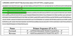
Figure 1. The particular target sequence of M. simiae, referred to as MST601, encompassing 601 base pairs. The locations and sequences of the forward and reverse primers are specified (Tm: 59.17 and 57.77, respectively; GC%: 50.00 for both).
|
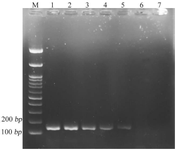
Figure 2. Agarose gel electrophoresis of MST601-PCR products using serial dilutions of M. simiae genomic DNA. M: 100 bp DNA marker, Lane 1: 100 pg, Lane 2: 10 pg, Lane 3: 1 pg, Lane 4: 100 fg, Lane 5: 10 fg, Lane 6: 5 fg, and Lane 7: 1 fg. A specific amplicon of 168 bp was detected up to the 10 fg (approximately equivalent to 1.5 copies of M. simiae genomic DNA per μl) with no amplification observed at 5 fg and 1 fg.
|
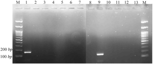
Figure 3. Specificity of MST601-PCR assessed using genomic DNA from various mycobacterial species. M: 100 bp DNA marker, Lane 1: positive control, Lane 2: M. bovis, Lane 3: M. smegmatis, Lane 4: M. kansasii, Lane 5: M. avium, Lane 6: M. chelonae, Lane 7: M. intracellulare, Lane 8: M. fortuitum, Lane 9: M. simiae, Lane 10: M. ulcerans, Lane 11: M. marinum, Lane 12: M. tuberculosis, and Lane 13: negative control. The absence of bands in non-simiae species confirms the high specificity of the assay, with no cross-reactivity observed.
|
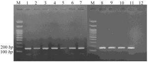
Figure 4. Agarose gel electrophoresis showing amplification of the 168 bp MST601 target in ten clinical isolates of M. simiae. Lane M: 100 bp DNA marker; Lane 1: positive control; Lanes 2-11: M. simiae isolates; and Lane 12: negative control.
|
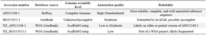
Table 1. Summary of the genome sequence records for M. simiae accessible in the NCBI database
The dependability and comprehensiveness of the entries exhibit considerable variation, with AP022568.1 serving as the fullest and most credible reference.
|
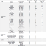
Table 2. Sequence alignment results of MST601 against various mycobacterial species
Query coverage is the proportion of the query sequence aligned with the reference genome, whereas identity signifies the percentage of identical nucleotides inside the aligned region.
|
|