Bioactive Silver Nanoparticles Coated with Curcumin Inhibit Angiogenesis through MMP-9 and Cox-2 Down-Regulation
-
 Ramezani Farzin , Tayebe
Farzanegan Campus of Semnan University, Semnan, Iran+98 2333464884 E-mail: tayeberamezani@semnan.ac.ir, Tel: +98 2333464884; E-mail: tayeberamezani@semnan.ac.ir
Ramezani Farzin , Tayebe
Farzanegan Campus of Semnan University, Semnan, Iran+98 2333464884 E-mail: tayeberamezani@semnan.ac.ir, Tel: +98 2333464884; E-mail: tayeberamezani@semnan.ac.ir
Abstract: Background: Inhibition of angiogenesis is an attractive approach in cancer therapy. Both curcumin and silver nanoparticles (AgNPs) have demonstrated anti-angiogenic properties; however, the the poor water solubility of Curcumin and the side effects of AgNPs adversely affect their activity.
Methods: In this study, AgNPs coated with curcumin (Cur-AgNPs), was used to improve aqueous-phase solubility of curcumin and decrease the side effects of AgNPs. After-wards, treatment with curcumin enhanced the anti-angiogenic activity of Cur-AgNPs. The nanoparticles were synthesized as both reducing and stabilizing agents. Evaluation of anti-angiogenesis was assessed in vitro using Human Umbilical Vein Endothelial Cells (HUVECs) and in vivo through the Chorioallantoic Membrane (CAM) assay. Da-ta were analyzed by one-way ANOVA with Tukey’s multiple comparison test.
Results: Synthesized Cur-AgNPs have an average diameter of 39 nm, with spherical shapes and an absorbance peak at 450 nm in the UV-visible spectrum. Cur-AgNPs showed a negative zeta potential. EDAX and FTIR confirmed the conjugation of cur-cumin with AgNPs. In vitro anti-angiogenesis assays demonstrated that Cur-AgNPs re-duced the viability of HUVECs, an Inhibitory Concentration (IC50) value of 13 µg/ml. DAPI and acridine orange/propidium iodide staining revealed a significant increase in apoptotic cells following treatment with Cur-AgNPs. The expression of Matrix Metallo-proteinase 9 (MMP-9) and Cyclooxygenase-2 (COX-2) was also inhibited in treated cells. In vivo anti-angiogenesis assays using the CAM model showed significant de-crease in the number, length and hemoglobin content of CAM blood vessels.
Conclusion: Curcumin conjugated with AgNPs may represent a promising strategy to enhance the therapeutic potential of both AgNPs and curcumin. However, further in-vestigations, particularly regarding safety and biocompatibility of Cur-AgNPs, is needed in this field.
Introduction :
Angiogenesis is a significant concept in cancer-related research, since the formation of new blood vessels is critical to the survival and metastasis of tumor cells. Anti-angiogenic therapy is one of the most critical strategies in combating cancer, as it inhibits or disrupts the growth of tumor blood vessels that provide essential nutrients and oxygen to maintain cancer cell activity 1. Due to the high mutation rate of tumor cells, they become resistant to anti-cancer drugs, while the low mutation rate of normal endothelial cells leads to their low resistance to such medications 2. Anti-angiogenesis agents are used in the treatment of some diseases that involve abnormal blood vessel growth under noncancerous conditions, such as macular degeneration 3. The FDA has approved certain drugs that have anti-angiogenic activity, including pazopanib, sorafenib, and sunitinib. Furthermore, the combination of angiogenesis inhibitors with additional therapies, especially chemotherapy, has reportedly increased their effectiveness 4.
Curcumin is an active ingredient of Curcuma longa with numerous pharmacological activities, such as anti-carcinogenic, anti-inflammatory, antioxidant, and anti-angiogenic properties. Curcumin functions as an angiogenic inhibitor by effectively suppressing several proangiogenic proteins, including Vascular Endothelial Growth Factor (VEGF) and basic Fibroblast Growth Factor (bFGF). It was also reported that curcumin reduces the viability of Human Umbilical Vein Endothelial Cells (HUVECs) 5,6. However, poor bioavailability of curcumin significantly limits its therapeutic activity, due to low absorption, rapid metabolism and fast elimination from the system 7,8. Researchers have attempted to both improve the solubility and targeted delivery of curcumin to cells. For instance, Zhang et al 9 developed a photoresponsive self-microemulsifying system carrying curcumin to improve both solubility of curcumin and its delivery to the colon 9. Ullah 10 formulated a photoactivated self-microemulsifying drug delivery system loaded with curcumin to provide a carrier for such hydrophobic molecules 10.
Recently, improving the bioavailability of curcumin using nanotechnology has become a significant concern among researchers. In the current work, an effort was made to increase the bioavailability of curcumin by designing a biological system. The medical properties of silver have been known for over 2,000 years 11. In recent years, anti-angiogenic and anti-cancerous effects of AgNPs have also been considered 12,13. This study aimed to develop curcumin coated silver nanoparticles (AgNPs) to decrease the side effects of AgNPs, enhancing nanoparticles stability and improving curcumin’s solubility, and bioavailability.
Materials and Methods :
Reagents: Curcumin (Sigma-Aldrich, United Kingdom), MTT [3-(4, 5-dimethylthiazol-2-yl)-2,5-diphenyltetrazolium bromide] (Sigma-Aldrich, United Kingdom), Acridine Orange (AO), and Ethidium Bromide (EB) (Sigma-Aldrich, United Kingdom), DAPI (4'-6-diamidino-2-phenylindole) (Sigma-Aldrich, United Kingdom), Annexin V/PI (Abcam, Germany), Caspase 9 assay kit (Abcam company, Germany), High Pure RNA Isolation Kit (Roche, Germany), cDNA Synthesis Kit (Fermentas, Germany), RPMI-1640 and and Fetal bovine serum (Invitrogen, United States). DMSO (dimethyl sulfoxide) (Merck, Germany), paraformaldehyde (Merck, Germany) and PCR kit (Parstous, Iran). HUVECs (Pastor cell bank, Iran), primers (Bioneers, Korea). All experiments were performed in at least three independent biological replicates.
Synthesis: The synthesis method of the Cur-AgNPs has been previously discribe by Ramezani et al in 2019 14. In the earlier work, their anti-cancer effects were evaluated, whereas in the present study, their anti-angiogenic properties were investigated. Briefly, Cur-AgNPs were synthesized by reducing an aqueous solution of AgNO₃ using curcumin as both the reducing and stabilizing agent. To this end, 500 ml of AgNO₃ (1 mM) was prepared, and 1 ml of curcumin (100 mM) was added to the solution and mixed thoroughly. The mixture was kept at room temperature until the color changed from yellow (curcumin) to brown, indicating the formation of Cur-AgNPs.
Characterization methods: The UV-visible absorbance spectra of Cur-AgNPs were recorded in the range of 300-700 nm using a spectrophotometer (Epoch Biotech, Winooski, VT, United States). The hydrodynamic size distribution and zeta potential of Cur-AgNPs were determined by Dynamic Light Scattering (DLS) at 25°C, using a medium viscosity of 0.89 cP, a fixed scattering angle of 90°, and a 678 nm, 90 mW laser (Cordovan, Vaso Particle, France). The morphology of the Cur-AgNPs was observed using Transmission Electron Microscopy (TEM, Hitachi, Tokyo, Japan). Energy- dispersive X-ray was performed to characterize the elemental composition (EDX, XL 30; Philips, Eindhoven, Netherlands).
In Vitro Anti-angiogenic Effects of Cur-AgNPs using Human umbilical vein endothelial cells: Cell culture: Human Umbilical Vein Endothelial Cells (HUVECs) were maintained in RPMI 1640 medium enriched with 10% FBS and 1% penicillin-streptomycin (100 U/ml). Cells were incubated at 37°C in a humidified environment containing 5% CO2.
Cytotoxicity assay: MTT Cytotoxicity assay: MTT Solution Preparation: A stock solution of MTT was prepared by dissolving 10 mg of MTT powder in 2 ml of Phosphate-Buffered Saline (PBS, pH=7.4). The solution was sterilized by passing through a 0.22 µm syringe filter and stored at 4°C in the dark. Prior to use, the MTT stock solution was equilibrated to room temperature.
HUVECs were cultured in T-25 flasks using complete culture medium until approximately 80% confluence was reached. The cells were then detached using trypsin-EDTA, centrifuged, and resuspended in fresh medium. Cell density was determined using a hemocytometer and trypan blue exclusion. The required cell number was calculated, and an appropriate volume was taken from the stock suspension. Cells were seeded in 96-well plates at a density of 1×10⁴ cells per well in 100 µl of complete medium. Each treatment condition was performed in triplicate. The plates were incubated overnight at 37°C with 5% CO₂ to allow cell attachment and recovery to logarithmic growth phase. After 24 hr, the medium was replaced with fresh complete medium containing Cur-AgNPs at final concentrations of 5, 10, 15, 20, and 40 µg/ml. The cells were incubated for another 24 hr under the same conditions. Following treatment, the medium was carefully removed, and 50 µl of fresh medium along with 5 µl of MTT solution (5 mg/ml in PBS) were added to each well. The plates were incubated at 37°C for 4 hr to allow the formation of insoluble formazan crystals. The supernatant was then discarded, and the formazan crystals were dissolved in 100 µl of DMSO per well. Absorbance was measured at 570 nm using a microplate reader (Epoch, Biotek, Winooski, VT, USA). Cell viability was expressed as a percentage of the untreated control using the formula:
Cell viability (%) = (A_treated / A_control) × 100.
Staining assays: AO/EB staining: HUVECs were cultured in 24-well plates at a density of 5×10⁴ cells/well. After 24 hr, the cells were treated with 13 µg/ml of Cur-AgNPs (corresponding to the IC₅₀) for 24 hr. Subsequently, the cells were harvested, centrifuged, and resuspended in an equal volume of AO-EB solution. The samples were then examined under a fluorescence microscope (Olympus, Tokyo, Japan). AO/EB staining quantification using ImageJ.
Treated and control cells were collected. The cells were then stained with a mixture of acridine orange and ethidium bromide according to procedure described above. The number of total and viable cells was counted using ImageJ software. The percentage of viable cells was calculated as follows:
% of viable cells=Number of viable cells nucleiTotal nuclei×100
At least three random microscopic fields per sample were analyzed, and the mean±Standard Deviation (SD) was reported. Statistical analysis was performed to compare the treatment groups with the control group.
DAPI staining: Endothelial cells were cultured in 24-well plates at a density of 5×10⁴ cells/well and treated with 13 µg/ml of Cur-AgNPs (corresponding to the IC₅₀) for 24 hr. The treated cells were then fixed with 4% paraformaldehyde, stained with DAPI solution (1 µg/ml) for 60 s, and observed under a fluorescence microscope (Olympus, Tokyo, Japan) using a 488 nm filter.
DAPI staining quantification using ImageJ: The total number of nuclei in microscopic images of the samples was counted. Apoptotic nuclei were identified based on their morphological features, including condensed and fragmented shapes. The percentage of viable cells was calculated using the following formula:
% of viable cells=Number ofcells nucleiTotal nuclei×100
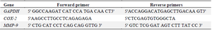
Gene expression assay by semi-quantified PCR: The IMPACT of Cur-AgNPs on the expression of Matrix Metalloproteinase 9 (MMP-9) and Cyclooxygenase-2 (COX-2) genes in HUVECs, was determined using the quantification Reverse Transcription Polymerase Chain Reaction (RT-PCR). The effect of Cur-AgNPs on the expression levels of MMP-9 and COX-2 genes in HUVECs was evaluated using quantitative Reverse Transcription Polymerase Chain Reaction (qRT-PCR). Endothelial cells were cultured at a density of 5×10⁶ cells 75 cm² flasks and treated with 13 µg/ml Cur-AgNPs for 24 hr. Total RNA was isolated and reverse-transcribed into complementary DNA (cDNA), which served as the template for PCR amplification with specific primers (Table 1). PCR reactions were carried out using a commercial kit (Parstuos, Iran) under the following conditions: initial denaturation at 94°C for 5 min, followed by 35 cycles of denaturation (94°C, 30 s), annealing (56°C, 30 s), and extension (68°C, 45 s), with a final elongation step at 72°C for 5 min. Amplified products were separated on 1.5% agarose gels and visualized using a gel documentation system (Unitec, Cambridge, UK). Band intensities were quantified using ImageJ software, and relative gene expression in treated cells was compared with that in untreated controls.
In-vivo assessment of ANT-angiogenic effects of Cur-AgNPs: Chick embryo chorioallantoic membrane assay: Fifty fertilized Ross chicken eggs were employed for the Chorioallantoic Membrane (CAM) assay and incubated at 37°C with 55-65% relative humidity throughout the experiment.
On the second day, a small window was created on each eggshell to access the CAM. On day 8 of incubation, in experimental groups, were treated with 10 µl of Cur-AgNPs at concentrations of 25, 50, 75, and 100 µg/ml. The treated eggs were then returned to the incubator. On day 12 of incubation, all eggs were examined and imaged using a stereomicroscope (Zeiss, Munich, Germany) equipped with a digital camera (Canon PowerShot G2).
Assessment of CAM vascularization and hemoglobin content: The vascularization network (number and length of blood vessels) surrounding the gelatin sponge was analyzed in all samples using ImageJ software. Additionally, the extent of vascularization was quantified based on the hemoglobin level in the CAM using the Drabkin method. Briefly, CAM tissues were homogenized for 15 min, and the homogenates were centrifuged at 5000 rpm for 5 min. The supernatant was mixed with 1 ml of Drabkin reagent. The absorbance of each sample was recorded at 546 nm using a spectrophotometer (Epoch Biotech, Winooski, VT, USA) according to the manufacturer’s instructions.
Ethical approval: This study was approved by the Ethics Committee of the Faculty of Veterinary Medicine, Semnan University, with the ethical code IR.SU.REC.1404.22.
Statistical analysis: All experiments were conducted in at least in triplicate, and data are presented as mean±Standard Deviation (SD). Statistical analyses were performed using one-way analysis of variance (ANOVA) followed by Tukey’s multiple comparison test. Differences were considered statistically significant at p<0.05.
Results :
Synthesis and characterization OF Cur-AgNPs: As previously reported, Cur-AgNPs were successfully fabricated using curcumin as a reducing and capping agent. UV-visible spectroscopy revealed a distinct absorbance peak at 440 nm, confirming the formation of AgNPs. Transmission Electron Microscopy (TEM) showed that Cur-AgNPs were mostly spherical in shape with a uniform distribution, while DLS analysis demonstrated an average nanoparticle size of 39 nm. The AgNPs exhibited a zeta potential in the negative range of -10 to -30 mV, indicating good colloidal stability. This negative surface charge generates repulsive forces between individual Cur-AgNPs, preventing agglomeration and contributing to their stability in solution. These findings indicate that the synthesis process produces well-formed and stable AgNPs suitable for various applications, depending on their size and morphology (Figures 1A and 1C).
The similarity between the FTIR spectral patterns of free curcumin and the Cur-AgNPs verified successful conjugation of curcumin on the surface of Cur-AgNPs due to presence of curcumin’s characteristic absorption bands in the FTIR spectra of the nanoparticles. This conjugation likely enhances the stability and physicochemical properties of the synthesized nanoparticles (Figure 1D). In addition, energy-dispersive X-ray spectroscopy was used to determine the elemental composition of Cur-AgNPs. The EDX spectra (Figure 1C) displayed prominent silver peaks, while the presence of oxygen and carbon peaks indicated successful conjugation of curcumin with the nanoparticles. These findings confirm the successful synthesis and interaction between curcumin and AgNPs (Figure 1E).
Anti-angiogenic effects and apoptosis induction of Cur-AgNPs on HUVECs:Cytotoxicity assay: The cytotoxic effects of Cur-AgNPs on HUVECs were evaluated using the MTT assay. The results demonstrated a clear dose-dependent decrease in cell viability, with an IC50 value of 13 µg/ml after 24 hr of treatment. Concentrations below 5 µg/ml did not significantly affect cell viability (p≥0.05) indicating that low concentrations of Cur-AgNPs is nontoxic for the cells while, concentrations above 40 µg/ml induced rapid and pronounced cytotoxicity, leading cell death within the first hour after treatment (p≤0.001; Figure 2). These finding suggest that Cur-AgNPs reduce endothelial cell viability and proliferation which may suppress new blood vessel formation. These results support the anti-angiogenesis properties of Cur-AgNPs.
Morphological change observation: DAPI staining: DAPI is a fluorescent dye with high affinity that binds to AT-based pairs in DNA. DAPI staining was used to evaluate nuclear morphology change in HUVECs following treatment with 13 µg/ml Cur-AgNPs for 24 hr. Compared to untreated groups, Cur-AgNPs-treated cells exhibited characteristic hallmarks of apoptosis, including chromatin condensation, cell shrinkage, and apoptotic body formation (Figures 3A and 3C). Quantitative analysis of DAPI-stained cells showed that 30% of cells (p=0.009) in the nanoparticle-treated groups exhibited signs of apoptosis. In contrast, cells in the untreated control group remained healthy, with cell viability around 95%. The apoptosis index measured by DAPI staining may appear lower than the decrease in viability observed in the MTT assay, as MTT reflects metabolic activity while DAPI detects nuclear changes associated with apoptosis. Furthermore, cells that detach during apoptosis or late necrosis are usually lost during the washing and staining process, and therefore are not counted under the microscope. These findings confirm that Cur-AgNPs induce apoptosis in endothelial cells, potentially contributing to their anti- angiogenesis activity.
AO/EB staining: AO/EB staining was used to assess the effects of 13 µg/ml Cur-AgNPs on HUVECs. In this assay cells exhibited green fluorescence, whereas apoptotic cells displayed bright orange to red nuclei due to chromatin condensation and DNA fragmentation. After 24 hr of exposure to 13 µg/ml of Cur-AgNPs, the number of apoptotic (orange) cells increased in Cur-AgNPs treated cells compared to the untreated control.
Quantitative analysis of AO/EB -stained cells showed that the percentage of viable cells in the treated group decreased by approximately 37%, and the presence of yellow and orange hues in the stained images indicated induction of apoptosis. In contrast, cells in the untreated control group mostly displayed green fluorescence, with cell viability remaining at about 98%. The Viale cells in treated groups reduced significs (p=0007). Representative fluorescence images clearly revealed apoptotic morphological features, including nuclear condensation and membrane blebbing in the treated cells. In this type of staining, as in DAPI staining, the percentage of dead cells may appear lower than that measured by the MTT assay. This is because cells in the late stages of cell death are often lost during the staining and washing procedures. Nevertheless, the relative increase in cell death in treated groups compared to the control is clearly observable. This result corroborates the DAPI staining data, supporting anti-angiogenic activity of Cur-AgNPs (Figure 2D and 2F).
Gene expression analysis: Semi-quantitative RT-PCR was employed to examine the expression of COX-2 and MMP-9 in HUVECs treated with 13 µg/ml of Cur-AgNPs. The results, indicated a significant downregulation MMP-9 expression in HUVECs (to 69% relative expression) compared to untreated control groups (p=0.005). Similarly, COX-2 expression decreased markedly (to 54% relative expression) compared to untreated controls groups (p=0.001; Figure 4). These finding suggests Cur-AgNPs induced apoptosis and modulate gene expression by suppressing key inflammatory and extracellular matrix degrading enzyme, thereby contributing to their anti-angiogenic potential.
In-vivo assessment of ANT-angiogenic effects of Cur-AgNPs: Chick embryo chorioallantoic membrane: The chick embryo chorioallantoic membrane model was utilized to evaluate the inhibitory potential of Cur-AgNPs on angiogenesis. The treatment resulted in a clear dose-dependent suppression of neovascularization, as reflected by the progressive reduction in both the mean vessel length and vessel density in treated sample compared with untreated controls.
Specifically, administration of 50 µg/ml and 100 µg/ml Cur-AgNPs significantly reduced the mean vessel length to 33 and 27 mm, respectively, compared with 40 mm in the control group (p=0.04 and p=0.002).
A similar trend was observed for vessel number, which decreased to an average of 40 and 33 vessels at 50 µg/ml and 100 µg/ml Cur-AgNPs, respectively, compared with 54 vessels in controls (p=0.002 and p<0.001). In contrast, treatment with 25 µg/ml Cur-AgNPs (mean vessel number=49; p=0.19) did not produce any significant alteration in vascularization, indicating a threshold concentration required for anti-angiogenic efficacy.
Evaluation of CAM vascularization and hemoglobin content: To further quantify vascular inhibition, hemoglobin content—an indirect indicator of vessel density—was determined using the Drabkin assay. A marked and statistically significant decrease in hemoglobin concentration was observed in CAMs treated with 50 µg/ml (mean=3.58) and 100 µg/ml (mean=2.50) Cur-AgNPs compared with untreated control (mean=3.90; p=0.006 and p=0.0001, respectively). The reduction in hemoglobin content was consistent with the observed decline in vessel number and length, suggesting effective vascular suppression (Figures 5 and 6). Collectively, these results provide compelling in vivo evidence of the potent anti-angiogenic activity of Cur-AgNPs. The findings imply that Cur-AgNPs effectively disrupt the formation and maturation of new blood vessels in a concentration-dependent manner, thereby supporting their potential.
Discussion :
AgNPs were synthesized through an environmentally friendly approach using pure curcumin as both a reducing and capping agent 14. The resulting nanoparticles exhibited a nearly spherical morphology with an average size of approximately 40 nm, as confirmed by TEM analysis, and displayed a negative zeta potential, indicating good colloidal stability. Fourier-transform infrared spectroscopy and energy-dispersive X-ray spectroscopy confirmed the successful conjugation of curcumin with AgNPs. Previous studies have reported the green synthesis of AgNPs using various plant extracts as reducing and stabilizing agents 15,16.
In the present study, pure curcumin was employed as a well-defined bioactive molecule for nanoparticle synthesis, in contrast to conventional approaches that rely on complex plant extracts. Similarly, Kabir et al 17 developed gold nanoparticles functionalized with silymarin, which acted as both a reducing and capping molecule, and the conjugation was verified using FTIR spectroscopy 17.
The inhibitory influence of Cur-AgNPs on angiogenesis was examined. The results revealed that Cur-AgNPs possess potent anti-angiogenic activity both in vivo and in vitro in a dose-dependent manner. Based on the findings from the CAM assay, Cur-AgNP treatment significantly reduced both the length and density of blood vessels on the chorioallantoic membrane. Furthermore, these nanoparticles markedly suppressed the proliferation of endothelial cells. To gain insight into the molecular mechanisms underlying this anti-angiogenic effect, the expression of COX-2 and MMP-9—two principal genes involved in angiogenesis and metastasis—was analyzed in Cur-AgNP–treated endothelial cells and compared with untreated controls. The expression of both MMP-9 and COX-2 was found to be substantially downregulated following exposure to Cur-AgNPs. Proteolytic enzymes belonging to the MMP family, particularly MMP-2 and MMP-9, are known to participate in extracellular matrix degradation, facilitating tumor invasion and vascular development. Consequently, inhibition of these enzymes can effectively limit endothelial cell migration and the formation of new blood vessel 18,19.
Consistent with the present findings, several previous studies have reported the inhibitory effects of curcumin on MMP-2, MMP-9, and COX-2 expression 20,21. Similarly, Gurunathan et al 22 demonstrated that AgNPs suppress angiogenesis by inhibiting the activation of the PI3K/Akt signaling pathway and MMP-2 22. In another investigation, Satapathy and colleagues developed hybrid AgNPs functionalized with quinacrine (QAgNPs) and assessed their anti-tumor potential in several oral cancer cell lines, including H-357, Oral Squamous Cell Carcinoma (OSCC), and Oral Cancer Stem Cells (OSCC-CSCs), using both in vitro and in vivo models. Their results revealed that QAgNPs exhibited superior cytotoxicity toward OSCC-CSCs and produced a more pronounced reduction in angiogenesis compared to either quinacrine or AgNPs alone 23.
Furthermore, Ajaykumar et al 24 evaluated the anti-angiogenic potential of AgNPs synthesized using Uvaria narum extract (Un-AgNPs) through a CAM assay, reporting a marked inhibition of neovascularization. These observations are in strong agreement with the results obtained in the present study. Despite the promising outcomes, this work has certain limitations. The current experiments were restricted to in vitro and in vivo settings, which, although informative, do not fully replicate complex physiological conditions. Therefore, additional studies employing animal models and, ultimately, clinical trials are necessary to evaluate the long-term safety, systemic distribution, and potential toxicity of Cur-AgNPs. Future investigations should also explore their performance in cancer models to assess their feasibility as targeted nanocarriers for site-specific drug delivery. Comparative analyses with conventional chemotherapeutic and anti-angiogenic agents could further clarify the therapeutic advantages and translational potential of these nanoparticles. Addressing these aspects will strengthen and extend the significance of the current findings.
On the same note, the inhibitory effects of curcumin on MMP-2, MMP-9, and COX-2 have been reported in previous studies 20,21. In this regard, research by Gurunathan et al demonstrated that AgNPs exert inhibitory effects on angiogenesis, noting that AgNPs could inhibit the activation of PI3K/Akt and MMP. In another study prepared hybrid AgNPs (Agnos) by molecule quinacrine (QC) and evaluated the anti-tumor activity of QAgNPs using various cell lines, including H-357 oral cancer, oral squamous cell carcinoma (OSCC) and Oral Cancer Stem Cells in in vivo and in vitro model systems. According to their findings, QAgNPs have greater ability in killing OSCC-CSCs compared to QC and AgNPs alone. Furthermore, they reported that QAgNPs reduced angiogenesis more significantly compared to QC and AgNPs 23,25. Ajaykumar et al 24 examianed the anti-angiogenic effects of AgNPs fabricated using Uvaria narum (Un-AgNps) extract.
For evaluating the anti-angiogenic effect of Un-AgNps the CMA was used. According to the results of this study, Un-AgNps show perfect anti-angiogenesis properties. These findings are consistent with the results of the present study 24.
Although this study provides valuable insights into the effects of AgNPs on HUVECs and CAMs, several limitations should be acknowledged. The current investigation was confined to in vitro and in vivo models, which, although informative, may not fully replicate the complexity of physiological conditions in living organisms. Therefore, future studies involving animal models and, ultimately, clinical trials are required to comprehensively assess the long-term safety, systemic distribution, and potential toxicity of AgNPs. Further investigations should also explore their behavior in tumor-bearing models to evaluate their potential as targeted nanocarriers for site-specific drug delivery. Moreover, comparative studies with conventional chemotherapeutic and anti-angiogenic agents would offer a more holistic understanding of the therapeutic potential of AgNPs. Addressing these aspects will help to validate and extend the current findings.
Conclusion :
In the present study, AgNPs were produced using pure curcumin as a non-ionic reducing agent. These nanoparticles were suitable in size for biological applications (39 nm) and also had good stability in aqueous environments (negative zeta potential). The results indicated that Cur-AgNPs induce apoptotic effects on HUVECs leading to inhibition of angiogenesis. Also in vivo CAM assay results confirm anti-angiogenesis properties of Cur-AgNPs. Overall, Cur-AgNPs show promise for future investigation in anti-angiogenesis and anti-cancer applications, although additional studies are required to fully understand therapeutic potential and safety.
Conflict of Interest :
The authors declare no conflict of interest.
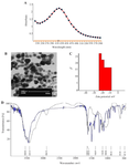
Figure 1. Synthesis and characterization of Cur-AgNPs.
A) UV–visible absorption spectrum of Cur-AgNPs. The absorption peak observed around 450 nm confirms the formation of silver nanoparticles.
B) TEM image of Cur-AgNPs showing spherical nanoparticles with good dispersion.
C) Zeta potential curve of Cur-AgNPs. The nanoparticles exhibited a negative potential ranging from ‐10 to ‐30 mV, which prevents their aggregation.
D) Comparison of FTIR spectra of curcumin (blue) and Cur-AgNPs (black). The similarity between the two spectra indicates that curcumin is bound to the silver nanoparticles.
|
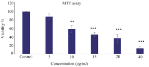
Figure 2. Viability assay by MTT assay. Cytotoxic effects of Cur-AgNPs on HUVECs after 24 hr. The results demonstrated that cell viability decreased in a dose-dependent manner in HUVECs treated with Cur-AgNPs compared with the control group. The data are presented as mean±SD (***p≤0.001).
|
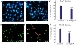
Figure 3. Cur-AgNPs (13 μg/ml) induced apoptosis in HUVECs after 24 hr.
A) Non-treated control group stained with DAPI. In this group, the cell nuclei remained intact, and no evidence of cell death was observed.
B) Cur-AgNP-treated group stained with DAPI. Evidence of apoptosis, such as nuclear condensation and disruption of cellular integrity, was observed.
C) Quantification of live cells in the treated sample compared with the untreated control. The graph shows an increased proportion of dead cells in the treated group compared with the control.
D) Control group stained with Acridine Orange/Ethidium Bromide (AO/EB), showing viable cells (green) with no signs of apoptosis.
E) Cur-AgNP-treated group stained with AO/EB. The treated cells exhibited apoptotic features (orange and green staining).
F) Quantification of live and dead cells in treated and control groups. The graph shows an increased proportion of dead cells in the treated group compared with the control. Arrows indicate apoptotic cells. Data are presented as mean±SD (magnification, ×200).
|
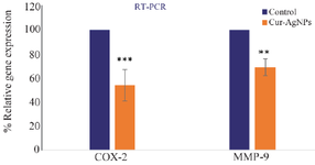
Figure 4. RT-PCR was performed to analyze the expression of COX-2 and MMP-9 genes in the control and the groups were treated by 13 µg/ml of Cur-AgNPs, (mean±SD), ***p≤0.001.
|
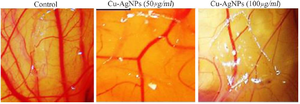
Figure 5. Average number and length of blood vessels in Chorioallantoic Membrane (CAM) samples were treated by different concentration of Cur-AgNPs compared to control.
|
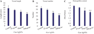
Figure 6. Effect of Cur-AgNPs on CAM angiogenesis.
A) Average length of CAM blood vessels in samples treated with different concentrations of Cur-AgNPs compared with the control. The results indicate a dose-dependent decrease in CAM blood vessel length. Treatment with 50 and 100 μg/ml Cur-AgNPs led to a significant reduction in vessel length compared with the control.
(B) Average number of CAM blood vessels in treated and control samples. A significant decrease in vessel number was observed at 50 and 100 μg/ml Cur-AgNPs compared with the control.
(C) Hemoglobin content measured using the Drabkin method. Samples treated with 100 μg/ml Cur-AgNPs showed a significant decrease in hemoglobin content relative to the control, indicating reduced angiogenesis.
Data are presented as mean±SD. *p≤0.05, p≤0.01, *p≤0.001.
|
|
|