Evaluation of the Anti-melanogenic Effect of Aqueous and Hydroalcoholic Extracts of Nasturtium officinale on the B16F10 Cell Line
-
Heydari , Mohaddese
-
Department of Pharmaceutical Biotechnology, School of Pharmacy and Pharmaceutical Sciences, Isfahan University of Medical Sciences, Isfahan, Iran
-
Sadeghi-Dinani , Masoud
-
Department of Pharmacognosy, School of Pharmacy and Pharmaceutical Sciences, Isfahan University of Medical Sciences, Isfahan, Iran
-
 Shafiee, Fatemeh
Department of Pharmaceutical Biotechnology, School of Pharmacy and Pharmaceutical Sciences, Isfahan University of Medical Sciences, Isfahan, Iran, Tel: +98 9133947450, 31 37922622; E-mail: f_shafiee@pharm.mui.ac.ir
Shafiee, Fatemeh
Department of Pharmaceutical Biotechnology, School of Pharmacy and Pharmaceutical Sciences, Isfahan University of Medical Sciences, Isfahan, Iran, Tel: +98 9133947450, 31 37922622; E-mail: f_shafiee@pharm.mui.ac.ir
Abstract: Background: This project aimed to evaluate the anti-melanogenic characteristics of Nasturtium officinale (N. officinale) by assessing the impact of both aqueous and hydroalcoholic extracts on the inhibition of cellular and mushroom tyrosinase enzymes, as well as the suppression of the melanin synthesis in B16F10 melanoma cells.
Methods: The aerial components of N. officinale were subjected to extraction using distilled water: ethanol (7:3) through the maceration technique. The extract’s phenolic compounds were quantified employing the Folin-Ciocalteu method. The evaluation of the safety profile of the extracts on B16F10 cells was done by the MTT assay. Subsequently, the melanin concentration in B16F10 cells, alongside the inhibitory effects on both mushroom and cellular tyrosinase, was assessed following treatment with the aforementioned extracts.
Results: The aqueous and hydroalcoholic extracts exhibited no significant toxicity on B16F10 when compared to Phosphate-Buffered Saline (PBS). Additionally, there was no notable difference in the cytotoxic effects of extracts on the B16F10 cell line. Both extracts resulted in inhibition of cellular and mushroom tyrosinase, along with a decrease in melanin levels in B16F10 in a concentration-dependent manner. Ultimately, the total phenolic content in the aqueous and hydroalcoholic extracts was found to be approximately 14 and 30 mg/g of gallic acid, respectively.
Conclusion: This in vitro investigation offers evidence supporting the skin brightening properties of N. officinale as an anti-melanogenic agent. Given its safety profile and absence of toxic effects on melanoma cells, it may be incorporated into the formulation of skin-brightening products following preclinical tests.
Introduction :
Hyperpigmentation refers to the darkening of a specific area of the skin or nails due to a rise in melanin levels 1. The management of hyperpigmentation through the use of skin-lightening cosmetic products, often applied topically, has received increasing attention in the public 2. The current skin-lightening agents on the market generally fall into one of the following categories in terms of their mechanisms of melanin inhibition: 1) targeted elimination of melanocytes, 2) suppression of melanosome development, 3) suppression of tyrosinase activity (an enzyme crucial in the conversion of tyrosine to melanin), 4) suppression of melanin production, and finally, 5) disruption of melanosome movement. Among these, tyrosinase inhibitors remain as the most prevalent skin-lightening agents with suitable specificity for targeting melanogenesis and good success 3.
Taking into account the adverse effects of specific chemicals that inhibit tyrosinase 4 and the higher preference for natural herbs among the public, it is vital to investigate and confirm the healing properties of herbs that have a history of traditional application. This method can aid in substituting chemical and entirely synthetic products with these alternatives. Furthermore, because of the resemblances in active compounds and their impacts among plant families, other plants belonging to the same family can be recognized for possible utilization 5. The majority of natural compounds currently applied as skin lightening agents in the market are tyrosinase inhibitors 3.
Nasturtium officinale (N. officinale), from the Brassicaceae family, is documented in the German Commission E Monograph (Phytotherapy) 6,7. This herb has been utilized in traditional medicine for managing hyperglycemia, hypertension, asthma, and cough. Recent research indicates that it possesses anti-cancer, antioxidant, antimicrobial, anti-inflammatory, anti-psoriatic, and cardioprotective effects 8.
On the other hand, N. officinale has been noted for its valuable effects in lowering glucose levels, detoxifying the blood, acting against scurvy (vitamin C deficiency), and tuberculosis. Finally, it acts as a diuretic, can be used as toothache relief, and serves as a laxative 8,9. It has been utilized for its antioxidant effects 10.
N. officinale is rich in tannins, terpenoids (such as carotenoids), polyphenols (flavonoids, phenolic acids, proanthocyanidins), glucosinolates, Phenylethylene Isothiocyanate (PEITC), and contains vitamins (B1, B2, B6, A, C, and E) along with minerals (magnesium, iron, phosphorus, iodine, copper, and calcium) 11,12.
Taking into account the anti-melanogenic activities and tyrosinase inhibitory characteristics of flavonoids found in different parts of N. officinale, along with its antioxidant properties and the presence of vitamins C and E, recognized for their skin-lightening ability (especially vitamin C) 9,13, and the historical application of this herbal medicine for hyperpigmentation treatment, this study aims to explore the effects of aqueous and hydroalcoholic extracts in vitro on murine melanoma cell line (B16F10). This study seeks to facilitate the possible advancement of the extracts as a medicinal product after additional preclinical and clinical investigations.
Materials and Methods :
Extraction of aerial part of N. officinale: The aerial parts of N. officinale were ground into a powder after being dried in the shade and sifted through a sieve of suitable mesh size. The obtained powder was subsequently utilized to create aqueous and hydroalcoholic extracts. To prepare the aqueous extract, 340 g of the dried powdered aerial parts were soaked and blended with 1.5 L of distilled water for 24 hr, utilizing the maceration technique. The blend was agitated for approximately 10 min using a mechanical stirrer, then filtered, and this process was repeated three times. The extracts were united and freeze-dried employing a lyophilizer. For the hydroalcoholic extract, the similar method was utilized with 400 mg of herb powder and a solvent blend of water: ethanol (3:7 ratio). Ultimately, the hydroalcoholic extract was evaporated using a rotary evaporator and a freeze dryer. The collected dry extracts were utilized to prepare various concentrations for testing 14.
Standardization of extracts: The Folin-Ciocalteau method was used to standardize the total phenolic content 14, using phenolic gallic acid in ethanol as the standard solution in various concentration. These solutions were used to draw the calibration curve of phenolic content of each sample. Next, 5.0 g of hydroalcoholic and aqueous extracts were dissolved separately in ethanol 96%. Exactly, 20 μl of each sample (standard, blank, and extracts samples solutions) were poured into small test tubes and 58.1 ml of distilled water was added to each sample. Then 100 μl of Folin-Ciocalteau reagent was added to the tubes and mixed. After 30 min, 30 μl of fresh sodium bicarbonate solution, was added to reach a final volume of 2 ml. The solution was kept for 2 hr at 20°C in a dark environment and the absorbance was read toward the blank at a wavelength of 765 nm and a standard curve was drawn 14.
Assessment of extracts safety on B16F10 cells: The B16F10 cell line and the MTT assay were utilized to assess the toxic effects of the extracts on cells. Cells were grown in 96-well plates at 5×10⁴ cells/ml concentration in DMEM high glucose culture medium and incubated for 24 hr. In the next day, all wells exposed to aqueous and hydroalcoholic extracts in logarithmic concentrations (0.5, 5, 50, 500, and 5000 µg/ml final concentration) in 20 µl final volume to identify the safe concentration range. Cell viability was evaluated after 48 hr through the MTT assay 15. Briefly, after this period of time, 20 µl of MTT solution (5 mg/ml) was added to all wells and incubated for 3 hr at 37°C. Then the supernatant of each well was discarded, and the well content (formazan crystals) was dissolved in 150 µl of DMSO. Finally, the absorbance reading was done via microplate reader in 570 nm wavelength. The cell viability percent was calculated for each concentration of various extracts using bellow equation.
Cell viability (%) = (A-B/C-B)×100, which A is the average OD570 value of the extracts in various concentrations, B is the average OD570 value of the null culture media (blank), and C is the average OD570 value of the negative control [Phosphate-Buffered Saline (PBS) treated cells].
Assessment of the suppressive effects of the extracts on mushroom tyrosinase enzyme: To evaluate the extract's inhibitory effect on mushroom tyrosinase, 25 µl of L-DOPA (0.5 mM), 70 ml of tyrosine (10 mM), and 875 µl of phosphate buffer were mixed together. Subsequently, 10 µl of the samples in various concentration (31.25, 62.5, 125, 250, and 500 µg/ml final concentration) was incorporated into the mixture. In the end, 10 µl of the tyrosinase enzyme (500 units/ml) was added, and the solution was well combined.
The mixture was incubated for 30 min at 37°C, and the quantity of dopachrome generated was assessed using spectrophotometry at 475 nm. The percentage of inhibition was subsequently determined using the equation below:
The percentage inhibition of tyrosinase activity (% is calculated as ((B-A)/B)×100, with B representing the average OD 475 value from the control and A being the average OD 475 value for various concentrations of two extracts 15.
Measurement of melanin level in cells: To assess melanin levels, 900 µl of B16F10 cells (5×10⁵ cells/ml) in DMEM culture media supplemented with 10% of Fetal Bovine Serum (FBS) and pen/ strep antibiotics, were initially grown in 24-well plates. After 24 hr, different concentrations of the extracts (31.25, 62.5, 125, 250, and 500 µg/ml final concentration) were added to the related wells. Following 48 hr of treatment, the cells were removed from the plate with trypsin, washed twice with PBS, and then lysed by adding 100 πL of 2 N NaOH, followed by a 30 min incubation at 100°C. The lysate underwent centrifugation at 16,000×g for 20 min, and the absorbance of the supernatant was recorded at 405 nm. The concentration of melanin was assessed by drawing a standard curve that shows melanin (purchased from Sigma, USA) absorbance across its various concentrations 13. Kojic acid, a known anti-pigmentation agent, was utilized as a positive control 15.
Evaluation of the inhibitory effects of extracts on cellular tyrosinase enzyme activity: To achieve this, B16F10 cells were grown in a 24-well plate at a density of 5×10⁵ cells/ml for a total final volume of 900 µl in DMEM culture medium. Treatment with various concentrations of the extracts (31.25, 62.5, 125, 250, and 500 µg/ml final concentration) was carried out after 24 hr. Two days post-treatment, the cells were lysed through the freeze-thaw technique (Alternating exposure to 37 and 20°C for 5 consecutive times) and by incorporating 1% Triton X-100. The lysate was subsequently centrifuged at 16,000×g for 20 min. Ultimately, 100 πL of the centrifuged supernatant was mixed with L-DOPA at a concentration of 2 mM, and the absorbance was recorded at 492 nm 15. Again, the percentage inhibition of tyrosinase activity [% was calculated as ((B-A)/B]×100, with B representing the average absorbance value from the negative control and A being the average absorbance value for various concentrations of two extracts 15.
Statistical analysis: Statistical analysis was conducted with SPSS 25 software. Analysis of variance (ANOVA) along with an appropriate post hoc test (Tukey’s) was utilized to identify the statistical differences among groups. Cells treated with PBS were considered as the negative control, while kojic acid served as the positive control in tyrosinase inhibition assay experiments. All experiments were conducted at least three times. A p<0.05 was considered the threshold for significance.
Results :
The extraction efficiency for the aqueous extract stands at 3.84% (w/w), whereas the hydroalcoholic extract has an efficiency of 4.7% (w/w). Based on the Folin-Ciocalteau method, following three repetitions, the polyphenol levels in the aqueous extract equate to 14 mg of gallic acid/g of dry extract, while the hydroalcoholic extract holds the equivalent of 30 mg of gallic acid/g of dry extract.
The MTT assay results indicated no notable difference in cytotoxic effects between the aqueous and hydroalcoholic extracts (p>0.05), except at the maximum concentration tested (5000 µg/ml). Actually, at this concentration (5000 µg/ml), the aqueous extract showed a notable cytotoxic impact when compared to the negative control (PBS) (p=0.00). Furthermore, the aqueous extract showed more toxicity than the hydroalcoholic extract (p=0.036). In comparison, the hydroalcoholic extract showed no notable cytotoxicity relative to the negative control at any examined concentration up to 5000 µg/ml (p>0.05) (Figure 1).
Based on the results of this part, the anti-melanogenic and tyrosinase inhibitory effects of the extracts in 50-500 µg/ml ranges were surveyed.
The analysis of data reveals that the inhibitory effects on mushroom tyrosinase for both extracts rise as the concentration increases. At the lowest concentration (31.25 µg/ml), two extracts showed no significant difference in their capacity to inhibit mushroom tyrosinase (p=0.42). At all other concentrations, a significant difference was noted in the effectiveness of two extracts in similar concentrations (p<0.05). Furthermore, regarding the comparison of the tyrosinase inhibitory effects of each extract to the negative control (water in exposure to the substrate and tyrosinase enzyme), only in 125 µg/ml and higher concentrations was the significant difference observed (p=0.016) (Figure 2).
A comparison of the inhibitory effects on cellular tyrosinase from two extracts, on the other hand, revealed no notable difference in the efficacy of both extracts (when compared to each other), except at the concentration of 62.25 µg/ml, where a significant difference was noted (p=0.042). In all other concentrations, the difference did not reach statistical significance (p>0.05). Regarding the comparison of tyrosinase inhibitory effects of each extract with the negative control (PBS treated cells), on the other hand, except in 31.25 µg/ml, with no significant difference (p=0.068), in the other concentration this difference was statistically significant for both extracts (p<0.05) (Figure 3).
Finally, the analysis of the melanin content test indicated that cells exposed to various concentrations of aqueous and hydroalcoholic extracts, along with cells subjected to PBS and kojic acid as negative and positive controls, respectively, displayed a reduction in melanin content as the extract concentration increased. Except for the concentration of 31.25 µg/ml, where there was a significant difference between the melanin content of cells encountered with the same concentration of each extract (p=0.03), no significant differences were observed between the two extracts in the other tested concentrations p>0.05). In comparison to the positive control, only the highest concentration of both extracts (500 µg/ml) showed the same effects. Finally, in comparison to the negative control, for both extracts and in all concentrations, there were significant differences (Figure 4).
Discussion :
The test results indicated that the MTT assay revealed no notable difference in the cytotoxic effects of both extracts, apart from the highest tested concentration (5000 µg/ml). Hence, it can be concluded that both extracts are comparably safe at concentrations under this threshold. The aqueous extract showed a notable cytotoxic impact only at 5000 µg/ml when compared to the negative control (PBS). Conversely, the hydroalcoholic extract exhibited no noteworthy cytotoxicity when compared to the negative control at any of the concentrations evaluated. The concentration found to be the safest for cell viability was 0.5 µg/ml. Similarly, research conducted by Nilash et al regarding the impact of N. officinale extract on oral cancer indicated that the MTT assay findings for the aqueous extract on human Oral Cancer Cells (OCC 24) and Healthy Fibroblast cells (HF2FF) revealed that cell viability was notably influenced by both concentration and duration of exposure. Cytotoxic effects were notably pronounced at 4 mg/ml and 8 mg/ml after 24 hr, whereas all concentrations under 0.5 mg/ml after 48 hr demonstrated statistically significant viability. This implies that the cytotoxic effects of the aqueous extract rise with higher concentrations 16. In this research, while no notable difference in toxicity was observed between two extracts at the maximum concentration examined, neither extract exhibited a significant difference when compared to the negative control. This suggests the safety of the analyzed extracts.
A different study conducted by Casanova et al explored the antigenotoxic effects of N. officinale against induced DNA damage in vivo, utilizing living cells from Swiss mice aged 4 to 8 weeks 17. The research revealed no notable difference between the negative control (0.9% NaCl) and cells exposed to the aqueous extract, irrespective of the dosage of N. officinale applied extract (0.5 or 1 mg/kg body weight). This similarity is probably a result of the doses being beneath toxic levels, and the research indicates that the extract might possess DNA-protective properties at concentrations below toxic limits.
When examining the inhibitory effects of hydroalcoholic and aqueous extracts of N. officinale on cellular tyrosinase, no statistically significant difference was observed between the two extracts regarding tyrosinase inhibition. Additionally, it was noted that as concentration rose, the inhibitory effects on mushroom tyrosinase increased for both extracts, except the lowest concentration (31.25 µg/ml), where no significant difference was found between the two extracts in terms of their ability to inhibit mushroom tyrosinase. In all other concentrations, a significant statistical difference was noted in the effects of two extract groups.
Several investigations have assessed different herbs for their ability to inhibit tyrosinase and their anti-melanogenic properties. For instance, Chang et al examined the ethanolic extracts from the roots and twigs of Morus alba (M. alba), noting tyrosinase inhibition rates that varied from 0 to 62% and from 0 to 78%, respectively, at concentrations ranging from 0 to 60 µg/ml. These effects are linked to the polyphenols found in the plant 18, which are also present in both M. alba and N. officinale, possibly clarifying its anti-melanogenic traits.
A different study in this area examined the tyrosinase inhibitory effects of several tropical plants, as indicated by Baurin et al. Among these, five plants demonstrated significant inhibitory effects (above 90%) on mushroom tyrosinase: Entada africana (94%), Portulaca pilosa (93%), Prosopis africana (91%), Stryphnodendron barbatimao (90%), and Cariniana brasiliensis (90%). M. alba, employed as a positive control, exhibited a 97% inhibitory effect on the activity of mushroom tyrosinase. The authors observed that while M. alba has considerable skin-lightening effects, its tyrosinase inhibitory effects were limited; its skin-lightening abilities were instead linked to hyaluronidase inhibition 19.
The current research indicated that the inhibitory effects of N. officinale extract from its aerial parts on tyrosinase enzyme activity were linked to the existence of several compounds such as vitamins E and C, flavonoids, and other antioxidants. At the maximum concentration evaluated (5000 µg/ml), the extract showed around 35% inhibition of mushroom tyrosinase and 60% reduction in cellular tyrosinase activity.
In the research conducted by Wang et al, the anti-melanogenic properties of Antrodia camphorata fruit extracts were assessed utilizing the identical methodology as in our investigation 20. They indicated an IC50 value of 1.25 mg/ml for the inhibition of mushroom tyrosinase activity by the alcoholic extract and an IC50 of 50 µg/ml for the inhibition of cellular tyrosinase activity. Conversely, our research revealed that the IC50 values for inhibiting both mushroom and cellular tyrosinase exceeded 5 mg/ml.
In this study, elevating the concentration of both aqueous and hydroalcoholic extracts generally led to a reduction in cellular melanin levels. There was no notable difference between the two extract groups, apart from a concentration of 31.25 µg/ml.
A comparable study conducted by Hwang and Lee 21 examined the inhibitory effects of various herbal extracts on tyrosinase activity, melanin production, and L-DOPA oxidation. Melanin concentrations were assessed in B16F10 murine cells, comparing 0.03 mM of kojic acid, eugenol, naringenin, protocatechuic acid, syringic acid, limonene, perillaldehyde, kaempferol, and resveratrol against a negative control. Given that kojic acid, the positive control in the present research, exhibited greater effectiveness than the majority of tested plants, it can be inferred that the extract of N. officinale, which inhibits melanin biosynthesis less than kojic acid, may still significantly lower melanin production in cells at comparable concentrations, in relation to the plants examined by Hwang and Lee 22. This effect in our instance is probably attributable to the presence of antioxidants, especially vitamin C, as shown in a study by Rigopoulos et al 22.
Conclusion :
Given the active compounds identified and the findings from this research, it can be concluded that the extracts from the aerial parts of N. officinale serve as valuable brightening agent for cosmetic applications. It has shown effects similar to standard kojic acid, though at greater concentrations, and has fewer side effects than current chemical agents. The inhibitory impact of hydroalcoholic and aqueous extracts from the aerial parts of N. officinale on the tyrosinase enzyme, along with the exploration of skin-lightening mechanisms, indicates that this effect is probably connected to flavonoids, the presence of vitamins E and C, and antioxidant characteristics. In this medicinal herb, the primary mechanism for skin-lightening is the suppression of the enzyme tyrosinase. Finally, given its safety profile and absence of toxic effects on melanoma cells, it may be incorporated into the formulation of skin-brightening products following preclinical tests.
Acknowledgement :
This project was approved by Ethics Committee of Isfahan University of Medical Sciences with ethics code IR.MUI.RESEARCH.REC.1400.498.
Funding: This paper was financially supported by Isfahan University of Medical Sciences (grant number: 3400951).
Conflict of Interest :
The authors declare no conflict of interest.

Figure 1. Cytotoxicity of aqueous and hydroalcoholic extracts of N. officinale against B16F10 cells.
* Represents the different toxicity between two types of extracts and the negative control. error bar represents SD.
|

Figure 2. Mushroom tyrosinase inhibitory effects of aqueous and hydroalcoholic extracts of N. officinale.
* Represents the different inhibitory effects between two types of extracts. Error bar represents SD.
|
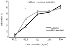
Figure 3. Cellular tyrosinase inhibitory effects of aqueous and hydroalcoholic extracts of N. officinale.
* Represents the different inhibitory effects between two types of extracts. Error bar represents SD.
|

Figure 4. Melanin content of cell extract treated with various concentrations of aqueous and hydroalcoholic extracts of N. officinale.
* Represents the different melanin content between two types of extracts. Error bar represents SD.
|
|