Potential Targets in Innate Immunity Receptors for Gastric Cancer: Insights from Virtual Screening in TCM and In Vitro Assay
-
Fakheri, Baratali
-
Department of Plant Breeding and Biotechnology, Faculty of Agriculture, University of Zabol, Zabol, Iran
-
 Bahari, Abbas
Department of Biotechnology, Research Institute of Modern Biological Techniques (RIMBT), University of Zanjan, Zanjan, Iran , Tel: +98 24 33054253; Fax: +98 24 33054254; E-mail: bahari@znu.ac.ir
Bahari, Abbas
Department of Biotechnology, Research Institute of Modern Biological Techniques (RIMBT), University of Zanjan, Zanjan, Iran , Tel: +98 24 33054253; Fax: +98 24 33054254; E-mail: bahari@znu.ac.ir
-
Fahmideh , Leila
-
Department of Plant Breeding and Biotechnology, Gorgan University of Agriculture Sciences and Natural Resources, Gorgan, Iran
-
Valadan, Reza
-
Department of Immunology, Molecular and Cell Biology Research Centre, Faculty of Medicine, Mazandaran University of Medical Sciences, Sari, Iran
-
Tavakolizadeh, Mahdi
-
Evidence-based Phytotherapy and Complementary Medicine Research Center, Alborz university of Medical Sciences, Karaj, Iran
-
Department of Pharmacognosy, School of Pharmacy, Zanjan University of Medical Sciences, Zanjan, Iran, Zanjan, Iran
Abstract: Background: Gastric Cancer (GC) poses a substantial global health threat, ranking as the second leading cause of cancer-related mortality among gastrointestinal malignancies. This investigation explores the potential therapeutic implications of plant extracts on gastric cancer, with a specific focus on their effects on the innate immune system.
Methods: A comprehensive analysis was conducted using 200 Sequence Read Runs (SRRs) thigh samples associated with gastrointestinal cancer tissue, juxtaposed with pathologically confirmed healthy tissues serving as controls. Differential Gene Expression (DGE) testing, encompassing the examination of 28,000 genes, including 95 pivotal genes associated with the innate immune system, was conducted. Findings elucidate alterations in the expression of key pattern recognition receptors, such as TLR2 and TLR4, as well as pivotal molecules within their signaling pathways. In pursuit of potential antagonists for these receptors, virtual screening on the Maestro docking platform in the Schrödinger 2022 package was conducted, evaluating 220,000 diverse tautomer’s of plant active substances. Selected candidates, exhibiting superior docking scores across four additional platforms, were subjected to further scrutiny.
Results: MMT results showed that nettle extract showed significant cytotoxic effects within 12 hr compared to the control (no treatment) sample, resulting in a 34.7% reduction in AGS cancer cell viability. The flow cytometry test showed that the control group had 71%, and groups treated with nettle extract for two and 12 hr had 65.3 and 67.18% viable cells, respectively. These differences were not statistically significant, indicating that nettle extract selectively preserves healthy living cells.
Conclusion: Cytotoxicity tests and cell cycle assessments confirmed the ability of nettle extract to reduce the survival of GC cells. This property makes nettle a promising candidate for drug development in this direction.
Introduction :
Cancer is the second leading cause of death in the world 1. Gastrointestinal cancers account for more than 25% of all cancer cases and more than 30% of cancer deaths worldwide 2. One significant factor that contributes to the development of cancer is inflammation. Infection is a known cause of inflammation, which is recognized by the innate immune system and leads to intense immune responses 3. The initial detection of infectious agents involves Pattern Recognition Receptors (PRRs) of the innate immune system, such as Toll-like Receptors (TLRs), RIG-I-like receptors, NOD-like receptors, and C-type lectin receptors 4. TLRs, in particular, function as a frontline defense against pathogens and can induce a range of inflammatory processes, influence cellular proliferation, and initiate apoptosis, all of which may facilitate tumor development. Among the various PRRs, TLRs have received the most attention in scientific research 5. Cytokines are pivotal in modulating inflammation and orchestrating immune responses. These secreted proteins not only coordinate host defense mechanisms but also play a central role in the onset and progression of immune-mediated diseases and cancers 6.
Understanding the molecular mechanisms and changes that precede the initiation and progression of gastric tumorigenesis is essential for early diagnosis and the identification of novel therapeutic and clinical targets for Gastric Cancer (GC). Various molecular abnormalities, including gene overexpression and gene silencing, have been identified in GC. However, elucidating the mechanisms underlying GC remains a significant challenge, as the molecular pathogenesis of GC is not yet fully understood. Gaining insights into these mechanisms could pave the way for more effective treatment strategies and enhanced patient outcomes by targeting specific pathways involved in the disease's development 7.
Researchers are exploring new therapeutic approaches for diseases alongside traditional treatments such as surgery, chemotherapy, and radiation therapy. One promising avenue is the use of in silico methods, which are both cost-effective and efficient for drug design and discovery 8. This study focuses on the potential therapeutic benefits of plant extracts for GC, specifically their impact on the innate immune system. Cell lines derived from gastrointestinal tissues, such as the human gastric adenocarcinoma (AGS) cells, are crucial for understanding the molecular mechanisms of gastric cancer and identifying potential treatments. Targeting these genes may help mitigate inflammatory pathways. Furthermore, virtual screening of the Traditional Chinese Medicine (TCM) library can uncover herbal compounds with the potential to antagonize pattern recognition receptors.
The present study explores the potential of extracts on this cell line and hypothesizes that among the approximately 100 key genes of the innate immune system, there are a number with significant differential expression in the transcriptome samples of gastrointestinal cancers that could be targeted to reduce inflammatory pathways, and virtual screening methods in the TCM library make it possible to identify herbal compounds that have antagonistic potential for pattern recognition receptors. Laboratory results show that compounds identified in selected medicinal plants are effective in reducing inflammation in gastrointestinal cancers.
Materials and Methods :
The methodology of this research was carried out in the following three stages:
1. One aim of the present study was to identify key receptors in inflammatory gastrointestinal cancers through transcriptomic analysis of at least 200 patients. First, 200 GC samples were sequenced using the SRA Fastq/SRA Toolkit package along with 200 healthy control samples in a Windows environment from the NCBI server. The data are derived from the transcriptomes of the samples themselves and are used as part of the tissue biopsy. The data were cleaned and trimmed for the presence of adapters and other sequencing elements, and then statistical analysis was performed for the DGE test in the cancerous and control groups. p-values less than one hundredth, absolute values, and fold changes greater than two were considered as significant increases or decreases. Therefore, 28,000 genes, especially 95 key genes of the innate immune system, were examined from this region 9.
2. Virtual screening was performed on the TCM database, and the molecular docking process was performed by Schrödinger 2022 software. In the next step, all leads that passed the docking stage (by Maestro) were docked by three software: CLC Drug Discovery, MOE, and ICM-Pro. The average scores from these programs were calculated and ranked based on the Schrödinger scores, leading to the selection of the highest-scoring compounds 10.
3. The leads filtered by virtual screening were searched in a previously prepared library of medicinal plants reported in the flora of Iran, and those pure compounds reported by the TCM library in a specific plant that were not present in the medicinal plants in the above library were removed from the list of final compounds 11. To confirm the results, in vitro analyses were performed on human Peripheral Blood Mononuclear Cells (PBMCs) cultured with the candidate plant extract, and changes in the expression of pro-inflammatory cytokines IL-1β and TNF-α (which play a role in inflammation, growth, support of cell proliferation, tumor regression, etc.) were examined by relevant experiments.
Statistical methods: Flow cytometric data for annexin PI were analyzed by determining cell counts at each cell stage, using ANOVA and Tukey's multiple-comparison test. The percentage of cells in each histogram region was calculated and reported using the FCS Express software on the flow cytometer.
Results an Discussion :
Library generation and ligand preparation: TCM has played an important role in drug discovery. The establishment of comprehensive TCM databases has significantly improved the efficiency and accuracy of research and made it easier to access information on TCM compounds 12. This library was prepared from a reputable traditional Chinese medicine database in Taiwan, and then virtual screening was performed on the TCM database according to the research objectives 13. Statistical analysis was performed for the DGE test in the two groups, cancerous and Ctrl. p values less than one hundredth, absolute values, and fold changes greater than two were considered as significant increases or decreases. Therefore, 28,000 genes, especially 95 key genes of the innate immune system, were screened from this region.
In the next step, several important genes in the inflammatory pathway were screened from the TCM database to determine the targets of anti-inflammatory drugs. All leads that successfully passed the initial screening stage by the maestro module. The Maestro module placed small ligand molecules within the receptor Glide grid and subjected them to three sequential docking steps, including High-Performance Virtual Screening (HTVS), Standard Precision (SP), and Extra Precision (XP). The time required to load each molecule and the scoring system used to evaluate each step differed from those of the previous steps. HTVS allows for rapid screening of many molecules in a limited space. XP docking, using a more refined scoring system, recognizes ligands with a Van der waals radius of 0.1 angstroms and a partial charge of 0.25. The program parameters were defined so that only 1 ligand state was retained per molecule (approximately 4.5% of the compounds remained).
In the secondary screening, their effects on the TLR4/MD2 complex were determined using three software programs: ICM, CLC Drug Discovery, and MOE, which were evaluated according to Lipinski's rules in BIOVIA Discovery Studio 2016. Figure 1 shows the four software used in this study. Also, Absorption, Distribution, Metabolism, Excretion-toxicity (ADME-Tox) were calculated using the Catalyst module in the above software, and additional compounds were removed. The quality of the obtained data was analyzed by FastQC software (https://www.bioinfor-matics.babraham.ac.uk/) 14. The average score across the three software programs was calculated, and the ranking was determined based on the average Schrödinger score. The chemical compounds and leads listed in table 1, selected from over 220,000 diverse tautomer’s, represent the most promising candidates for influencing TLR4. These compounds can either inhibit or activate these receptors.
Urtica dioica was selected from among the medicinal plants obtained in the first stage of the experiment through transcriptome analysis and examination of their active compounds as Toll-like receptor inhibitors. Additionally, due to its high ranking score by four software programs in primary and secondary screening, and its availability in the Iranian Flora Library after hydroalcoholic extraction of the plant, its effects on AGS cell line were investigated in cell tissue culture medium, and cell survival was measured with MTT (Methyl Thiazol Tetrazolium). Also, Real-Time PCR and flow cytometry (Annexin PI) experiments were performed in human peripheral blood mononuclear cells culture.
Cell culture and laboratory confirmation: To validate the results of bioinformatics studies, the assessment of inflammatory characteristics in in vitro cell culture will be performed as follows: The passage was started when the cell culture reached about 80% confluence. Also, the AGS cell culture under the microscope indicated that the cells were viable and in good numbers for testing (Figure 2). The AGS cell line was purchased from the Pasteur Institute Cell Bank (Tehran, Iran) (Table 2).
Preparation of hydroalcoholic extract of Urtica dioica: The plant leaves were stored in a dark and dry environment for the extraction process. To prepare a hydroalcoholic extract of nettle plant, 50 g of nettle leaf powder was soaked in 1000 ml of 70% ethanol, and the solvent-to-plant ratio was 20:1 15.
MTT assay: The use of cell culture methods provides a wealth of information regarding the effects of medicinal plant extracts on both cancer and normal cells. One method for measuring cell viability is the MTT assay. Cells treated with nettle hydroalcoholic extract (one mg of total extract) were examined for cell viability by the MTT technique after 2 and 12 hr. To prepare the MTT solution with a concentration of 5 mg/ml, 50 mg of MTT powder was dissolved in 10 ml of 0.15 M Phosphate-Buffered Saline (PBS) and diluted 10 times with sterile PBS during staining to obtain a 50 mg/ml solution of MTT. Then, 100 μl of Dimethyl Sulfoxide (DMSO) from Merck, Germany, was added to each well of the plate, and after 15 min of incubation at room temperature, their optical absorption was finally measured by Enzyme-Linked Immunosorbent Assay (ELISA) at a wavelength of 570 nm. To obtain better results and ensure the accuracy of the results obtained, each of the experiments was repeated three times 16.
The percentage of cell viability in cells treated for 2 hr was not significant compared to the control group, and this index was significant in cells treated for 12 hr, indicating the toxicity of the extract on cancer cells. Increasing the treatment time with the hydroalcoholic extract of the nettle plant reduces the survival of the AGS cell line (Figure 3).
Cell viability and apoptosis assay: To determine the level of apoptosis, an Annexin-PI (Propidium Iodide) kit in conjunction with flow cytometry was utilized. This approach allowed the researchers to investigate cell death and differentiate between early and late apoptosis, as well as necrosis. Cells were stained with the PI kit according to the manufacturer’s instructions. In this study, FCS Express 7 software was used to analyze the results obtained from flow cytometry. Also the types of induced cell death, specifically apoptosis and necrosis, in both control PBMC cells and those treated with nettle extract for 2 and 12 hr was assessed. The two reagents used in this technique were annexin V, which indicates apoptosis, and PI, which indicates necrosis figures 4 and 5.
It is fascinating to learn that PI cannot be absorbed by living cells, nor can it enter a healthy cell. However, when the cell membrane becomes necrotic, the permeability for PI increases, allowing PI to enter the cell. The PI absorbs a wavelength and emits a wavelength that can be read by flow cytometry. On the other hand, Annexin V can enter the cell, but it binds to or affects enzymes released from mitochondrial disintegration during the initial stage of apoptosis. This dye also absorbs and emits a specific wavelength, which can be read by flow cytometry. By using both dyes, it is possible to determine whether the cell is undergoing necrosis or apoptosis. If the cell only absorbs PI, it indicates necrosis, while absorption of only Annexin V indicates apoptosis.
Results of the PI test conducted in this study. The percentage of viable cells in the first group or control sample was 71%. In comparison, the percentage of viable cells in the second group, which was treated with nettle extract for two hr, was 65.3%. The third group, which was treated with the plant extract for 12 hr, showed a percentage of viable cells of 67.18%. These results indicate that nettle extract acts selectively and does not harm healthy living cells, and further research in animal models will be very useful to determine the effectiveness of nettle extract in the treatment of cancer.
Gene expression: RNA evaluation was conducted using a Nanodrop spectrophotometer. Each absorption unit at 261 nm equated to 41 ng/μl of RNA. The absorbance ratios at 261 nm to 231 nm and 211 nm indicated possible contamination with phenolic compounds and proteins, respectively.
Primer design: The primers used in the reaction were designed according to table 3. The housekeeping gene GAPDH (Glyceraldehyde 3-phosphate dehydrogenase) was chosen as the internal control or reference gene.
Quantitative PCR reaction: To amplify cDNA and conduct RT-PCR, a ROTOGEN 6000 device along with the SYBR Green qPCR MasterMix 5X kit from Pishgam Company was utilized. Materials required to perform the qPCR reaction are in table 4. The melting curve in figure 6 shows a single peak for each gene, indicating successful amplification of the target fragments and optimal PCR efficiency. The melting point also confirms the specific amplification of the target genes. Amplification curves for these genes were repeated in triplicate for each sample to ensure the accuracy and consistency of the results.
As the transcription factor NF-κB is located downstream of the TLR4 signaling pathway, its stimulation should result in an increase in the expression of proinflammatory cytokines such as IL1β and TNFα. Therefore, the expression of these two important genes in the TLR4 pathway in PBMC was measured using RT-PCR (Figure 7). Upon activation by its ligand, TLR4 triggers the activation of NF-κB, which then translocates from the cytoplasm to the nucleus, where it upregulates the expression of various genes, including IL-1β and TNFα, known as markers of inflammation. This study hypothesized that treating the cells with the extract would decrease the expression of these two genes; however, this was not observed.
The results showed that nettle extract has cytotoxic effects on AGS cancer cells in vitro. Soltani et al highlighted the anticancer potential of nettle, noting that its cytotoxic effects increase proportionally with higher extract concentrations 15. In the present study, we observed that exposing AGS cancer cells to a hydroalcoholic nettle extract for both 2 and 12 hr resulted in marked cytotoxicity and suppression of cell proliferation. Specifically, the most pronounced effects were observed after 12 hr of treatment, indicating the cytotoxic activity of the plant extract in this experiment. Furthermore, results from the Annexin PI assay demonstrated that the viability of human PBMCs remained unaffected by the treatment, suggesting that the nettle extract selectively targets cancerous cells while sparing normal cells. Cytokines are protein molecules that act as messengers in the circulatory system, transmitting immune system messages. Two of the most well-known of these are IL-1β and TNF-α, which are known as pro-inflammatory cytokines. When TLR4 is activated by its ligand, it leads to the activation of the transcription factor NF-κB. This factor then moves from the cytoplasm into the nucleus, where it increases the expression of genes, including IL-1β and TNF-α, which are known as markers of inflammation. The study hypothesized that treating the cells with the extract would decrease the expression of these two genes. However, this was not observed. One possible explanation for this is that PBMC cells are a mixture of lymphocytes and monocytes, which have different behaviors and gene expression. Additionally, PBMC cells are not fully functional, as T cells and B cells are immature, and monocytes have not yet differentiated into macrophages.
Therefore, researchers suggest a more detailed study on the identification of chemical compounds in the nettle plant. A complete understanding of the medicinal properties of this plant will make it a suitable species for medicinal and food uses. The anticancer effects of the nettle plant can be studied in animal models (in vivo) so that the compounds of this plant can be used more accurately in drug design. In this study, the treatment of target cells with nettle plant extract was performed at 2 and 12 hr, and based on the results, dynamic experiments can be performed at different times and concentrations to better understand the mechanism.
Conclusion :
The results of the present study, cytotoxicity tests and cell cycle assessments have demonstrated the efficacy of nettle extract in reducing the viability of GC cells. This study aimed to investigate the hypothesis that nettle, particularly its active compound fumaric acid, along with other substances traditionally recognized for their anti-inflammatory properties, could play a role in mitigating inflammation. However, the findings did not indicate a significant reduction in two specific inflammation indicators concerning gene expression. One potential explanation for this observation may be attributed to the use of PBMC cells, which comprise a mixture of various cell types. Future studies should focus on evaluating monocytes and lymphocytes separately to enhance the understanding of the effects observed.
Acknowledgement :
We sincerely thank and appreciate our colleagues and friends at the Universities of Zabol and Zanjan for their assistance in this work. This research is one of the articles resulting from the thesis of Mr. Abbas Ganjali, a doctoral student in the field of Agricultural Biotechnology at the University of Zabol, entitled "Screening of medicinal plants in the treatment of innate immune failures in GC: Insights from transcriptome analysis, virtual screening, and cell culture".
Conflict of Interest :
The authors declare that they have no conflicts of interest for the present article.
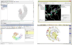
Figure 1. Four software used in the study: ICM, CLC drug discovery, maestro, and MOE.
|
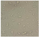
Figure 2. Confluency of AGS cell line under microscope at 40 magnification. AGS cells were cultured in RPMI 1640 medium supplemented with 10% fetal bovine serum (FBS) and 1% penicillin/streptomycin (100 U/ml) to inhibit fungal growth. The cells were maintained in an incubator under conditions of 5% CO2, 90% humidity, and 37°C.
|
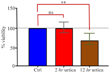
Figure 3. MTT Test results.
ns (not significant): indicates non-significance, * and ** indicate significance levels of 5 and 1%, respectively.
|
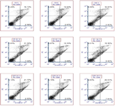
Figure 4. Histogram of flow cytometry analysis results in PBMC (Peripheral blood mononuclear cells) in triplicate for control and treatment samples at 2 and 12 hr.
|
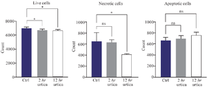
Figure 5. Apoptotic and necrotic changes in Peripheral Blood Mononuclear Cells (PBMC) treated with nettle extract.
ns (not significant): indicates non-significance, and * and ** indicate significance levels of 5 and 1%, respectively.
|
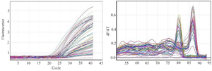
Figure 6. Derivative melting curve per unit time and fluorescence curve for two target genes.
The melting curve shows a single peak for each gene, indicating successful amplification of the target fragments and optimal PCR efficiency (Right image). The presence of parallel curves in the fluorescence diagram indicates the amplification of fragments in all samples (Left image).
|
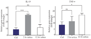
Figure 7. Expression of pro-inflammatory cytokines *, **, and *** indicate p-values less than 0.5, 0.01, and 0.001, respectively.
|
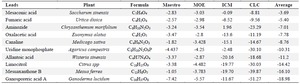
Table 1. Virtual screening by software (primary and secondary screening)
The plant compounds and sources are ranked based on the best average scores in four separate docking processes.
|

Table 2. Characteristics of AGS cell line
|
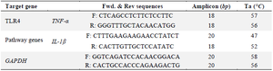
Table 3. Primers for gene expression reactions
|
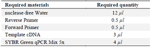
Table 4. Materials required to perform the qPCR reaction
The Real-time PCR experiment was conducted with two technical replicates. To minimize errors and enhance efficiency, a master mix containing primers, 5x SYBR Green qPCR Mix, and deionized water was prepared in a microtube and thoroughly mixed. After preparing the master mix, the appropriate amount was pipetted into each microtube, followed by the addition of the cDNA sample.
|
|