CpG-Containing Oligodeoxynucleotides and Freund Adjuvant in Combination with Alum Augment the Production of Monoclonal Antibodies Against Recombinant HBsAg
-
Khayyati Kohnehshahri, Mahsa
-
Department of Microbiology, Faculty of Veterinary Medicine, Urmia University, Urmia, Iran
-
Department of Cellular and Molecular Biotechnology, Institute of Biotechnology, Urmia University, Urmia, Iran
-
 Delirezh , Nowruz
Department of Microbiology, Faculty of Veterinary Medicine, Urmia University, n.delirezh@urmia.ac.ir, delirezhn@gmail.com
Delirezh , Nowruz
Department of Microbiology, Faculty of Veterinary Medicine, Urmia University, n.delirezh@urmia.ac.ir, delirezhn@gmail.com
-
Department of Microbiology, Faculty of Veterinary Medicine, Urmia University, Urmia, Iran
-
Department of Cellular and Molecular Biotechnology, Institute of Biotechnology, Urmia University, Urmia, Iran
Abstract: Background: Adjuvants are essential to potentiate the immune response to inoculated antigens and play a central role in vaccine development. Alum is generally used as a classic adjuvant, although it does not stimulate proper immunity, and some of the immunized subjects have low or no antibody response. Efforts have been continued to find more efficient adjuvants for better antibody responses. In the present study, the efficacy of three formulations of adjuvants, i.e. Cysteine p Guanine Oligodeoxynucleotide (CpG ODN), alum, and Freund, in the production of monoclonal anti Hepatitis B Surface Antigen (HBsAg) antibodies was investigated.
Methods: To immunize mice, regular hepatitis B vaccine containing recombinant HBsAg and alum was used with CpG ODN or Freund adjuvants, and splenocytes of hyperimmunized mice were fused with murine myeloma Sp2/0 cells. Positive hybridomas were selected by Enzyme-Linked Immunosorbent Assay (ELISA) using HBsAg as coating antigen followed by a limited dilution process.
Results: The results showed that by using all three formulations of adjuvants, monoclonal antibody (mAb) specific to HBsAg was successfully generated. It was also found that the mice immunized with (HBsAg + Alum) + CpG had the highest concentration of antibody production in serum and hybridoma supernatants as well as positive clones. Based on these findings, the addition of CpG ODN also induced a higher antibody response compared with Complete Freund’s Adjuvant (CFA).
Conclusion: Results of this study showed that CpG and Freund adjuvants could be efficient partners for alum in the immunization period of the process of monoclonal antibody production.
Introduction :
Serum includes various types of antibodies (polyclonal) which are particular to many types of antigens. Using a combined population of these antibodies causes different problems in immunochemistry techniques. Accordingly, obtaining homogeneous or monoclonal antibodies with defined preparation procedures has long been a goal for immunochemistry researchers, which could be achieved by hybridoma production technique. For the first time, Milstein and Kohler in 1975 developed a technique which helped to fuse a myeloma cell with an antibody secreting cell from an immunized animal and thus produced a hybridoma. These hybrid cells or hybridomas could survive in the laboratory and continue the secretion of particular antibodies. Antibodies secreted from hybridomas are
known as monoclonal antibodies 1,2. Any particle capable of stimulating humoral immune responses could be used for monoclonal antibody preparation. This particle could be a protein, carbohydrate, or nucleic acid. Monoclonal antibodies are used for cell staining, immunoprecipitation, immunoblot, immunoaffinity purification, and immunoassays with antigens or antibodies 3.
By fusion between suitable myeloma cells and spleen cells of the immunized animal, hybridoma could be obtained, which is a prerequisite for particular monoclonal antibody production objectives. Myeloma cells provide the genes which cause nonstop division in tissue culture, while antibody-secreting cells provide genes of immunoglobulins 1.
Of course immunogenicity of a vaccine is related to multiple factors including environmental, behavioral and nutritional factors, as well as vaccine factors such as administration route, vaccine dose and adjuvants. When the antigen is poorly immunogenic, the immune system needs an effective immune response inducer. Adjuvants capable of leading the responses towards cellular or humoral responses could be used for this purpose 2. In this study, three common adjutants, alum, Freund, and CPG ODN, were compared. Alum has been used for the past 70 years because of its immunostimulating potency. It is considered that alum increases the uptake of antigens from the injection site, which helps Antigen Presenting Cells (APCs) present the antigen efficiently 3. Alum can usually stimulate Th2 but not Th1; however, 10% of individuals do not possess the capability of antibody production with alum 4.
Seeking more effective adjutants which could stimulate Th1 and induce antibody production with high titer has shown that bacterial DNA, and not vertebral DNA, has immune stimulatory effects on leukocytes in vitro 5,6. This ability of bacterial DNA is because of unmethylated CpG dinucleotides, which are methylated in vertebrates 7. Leukocytes activation may also occur by artificial Oligodeoxynucleotides (ODNs) which have one or more CpG dinucleotides 7. Perhaps, the fast activation of the immune system in response to CpG ODN is due to the mechanisms of the innate immune system of vertebrates against Pathogen-Associat-ed Molecular Patterns (PAMPs) in micro-organisms 8. CpG ODN can induce proliferation in all types of B cells, which leads to secretion of polyclonal immunoglobulins 9-12. CpG ODN also causes improvement in procedures that induce Th1 cytokines, including IL-12 and IFN-γ, as well as those that induce the production of CTL and increase its activity. Moreover, it seemingly induces Th2 cytokines 10.
The Freund adjuvant, as the most powerful immunostimulatory reagent, has two types: Complete Freund Adjuvant (CFA) and Incomplete Freund Adjuvant (IFA) 13. These types of adjuvants exert their effects by affecting dendritic cells which induce stimulation and differentiation in T cells 14. The only significant concern about CFA is inflammation in the site of the injection which causes injuries 15.
Hepatitis B is one of the most common infectious diseases in the world. Hepatitis B virus (HBV) produces 4 major antigens, HBcAg, HBeAg, HBxAg, and HBsAg, which are related to the life cycle of the virus. Among them, the most important one is Hepatitis B Surface Antigen (HBsAg) which is found on the surface of virus and has attracted the attention of researchers for vaccine production 16. HBV can spread through the body fluids of the infected person, and human is the only natural host of this virus 17,18. It is known that HBV has three routes of transmission: perinatal, sexual, and parenteral 19,20. Interestingly, HBV has geographical prevalence. Three main strategies have been developed to prevent HBV infection, which include behavior changing, inactive immunoprophylaxis, and active immunization 21.
Because of the multiplex effects of different adjuvants in stimulating various aspects of immune responses and considering the production of the highest level of antibodies in immunized mice, the present study applied different combinations of CpG ODN, alum, and Freund adjuvants to immunize Balb/c mice and produce monoclonal antibodies against HBsAg.
Materials and Methods :
Mice
Inbred female Balb/c mice 6-8 weeks of age were purchased from the animal breeding department of Pasteur Institute of Iran (Pasteur Institute, Tehran, Iran). All mice were kept in plastic cages that were changed twice a week. Mice were maintained under approved conditions of a 12 hr dark/12 hr light cycle, at room temperature (22±2°C), and relative humidity of 55±5% and fed a standard diet and water. All experiments detailed here were approved by the Animal Research Ethics Committee of the University of Urmia (Ref No: IR-UU-AEC-138/DA/3), following the guidelines for the care and use of laboratory animals (National Institutes of Health, publication No. 8023, revised 1978).
Experimental groups
Twelve mice were randomly divided into 4 groups containing 3 mice each. Hepatitis B vaccine from Pasteur Institute of Iran (IRC 1228070570), which contained recombinant Hepatitis B Surface Antigen (rHBs-Ag) as immunogen and aluminum potassium sulfate (alum) as adjuvant was injected into three mice in group A. In group B, CFA (Sigma–Aldrich Co. Germany) was added to HBsAg plus alum. In group C, the mixture of HBsAg plus alum and oligodeoxynucleotides containing CpG motifs (CpG-ODN) (Invitrogen Co. USA) was used to immunize the mice, and the fourth group (D) were injected with Phosphate-Buffer-ed Saline (PBS) to serve as control. Therefore 4 groups were (HBsAg+Alum), (HBsAg+Alum)+FA, (HBsAg+ Alum)+CpG and control groups 22,23.
Immunization
Each mouse was intraperitoneally immunized 4 times at intervals of 3 weeks. The first immunization was performed using 50 μg of HBsAg and alum in 500 μl of vaccine preparation (group A), vaccine plus CFA (group B), and vaccine plus CpG ODN (10 μg). In the second and third rounds of immunization vaccine preparation, vaccine plus incomplete Freund adjuvant and vaccine plus CpG ODN (10 μg) were injected into mice in groups A, B, and C, respectively. In the last injection, mice in all three groups received the vaccine alone (without any Freund adjuvant or CpG ODN) as a booster dose three days before cell fusion (Table 1).
One week after the third immunization, blood samples were collected by a vertical incision of the tail vein of mice for determining antibody titers by indirect Enzyme Linked Immunosorbent Assay (indirect ELISA). Sera collected from non-immunized and immunized mice served as negative and positive controls, respectively. Efficiently immunized mice were determined to continue the process of monoclonal antibody production 23-25.
Indirect ELISA
Microtiter plates (Sigma-Aldrich Cat#CLS3590) were coated with the HBsAg (Pasteur Institute of Iran, Tehran, Iran) by adding 100 μl of protein solution in a coating buffer (50 mM Na2CO3/NaCO3H, pH=9.6) and incubated overnight at 4°C. Plates were blocked for 30 min at Room Temperature (RT) in 200 μl of 1% Bovine Serum Albumin (BSA) in PBS-Tween (0.05%) and then incubated with diluted serum samples (dilution 1:1000) for 2 hr at 37°C. After washing, the plates were incubated with 100 μl (dilution 1:1000) of anti-mouse antibody coupled with HRP diluted in 0.05% PBS (Sigma-Aldrich Co. Germany) and incubated for 2 hr at 37°C. The enzymatic reaction was developed using Tetramethylbenzidine (TMB) (Sigma-Aldrich Co. Germany) and terminated with 8.3% 12 mol/L HCl. The optical density was measured at 450 nm in a microplate reader (Biotec, USA).
Sp2/0 cell culture
Sp2/0 murine myeloma cell line was purchased from the National Cell Bank of Iran (NCBI C604, Pasteur Institute, Tehran, Iran) and cultured in RPMI 1640, 100 U/ml penicillin, 100 µg/ml streptomycin (Sigma-Aldrich Co. Germany), 15% FBS (Gibco, USA), and 5% CO2 at 37oC for 10-14 days to reach log phase and obtain 80% confluency and more than 95% viability which could be regarded as suitable for fusion.
Splenic cell preparation
The spleen was removed and cells suspension were prepared and transferred to a 15 ml conical and spun at 800 g for 3 min. The supernatant was discarded and the cell pellet resuspended in 1 ml lysis buffer, incubated at RT for 5-10 min. Then 9 ml RPMI-1640 was added and the mixture spun as before. The supernatant was then discarded and the cell pellet resuspended in 3 ml RPMI-1640. Cell count and viability were determined using trypan blue (Sigma-Aldrich Co. Germany) staining, and the resultant immunized mouse splenic cells which contained antibody producing B cells were used to make fusion with the Sp2/0 myeloma cell line 26,27.
Feeder layer
To collect murine peritoneal macrophages, mice were euthanized and the abdomen of each mouse wetted with 70% alcohol to sterilize the area. Ten ml RPMI 1640 medium was injected into the peritoneum of each mouse and fluid collected using a single 20-ml syringe. The needle was removed from syringe and the peritoneal fluid dispensed into 15-ml polypropylene centrifuge tubes on ice. Peritoneal lavage fluid in 15 ml tubes were centrifuged for 10 min at 400 g and 4°C. Then the supernatant was discarded and the cell pellet resuspended and adjusted to the appropriate cell concentration in RPMI 1640 medium to transfer into 96-well microtiter plates as the feeder layer.
Cell fusion
Sp2/0 cell line and splenic cells fusion was carried out three days after final immunization. The myeloma partner of fusion, which was prepared 10-14 days before fusion, reached log phase and was regarded as suitable for fusion with splenic cells at a ratio of 1:5 (1 SP2/0 and 5 spleen cells) by polyethyleneglycol (PEG, MW 4000) (Sigma-Aldrich Co. Germany) as fusion mediator. Medium containing Hypoxanthine, Aminopterin, and Thymidine (HAT) (Sigma-Aldrich Co. Germany) was added to the fused cells, and the cells were seeded into 96-well microtiter plates containing murine peritoneal derived macrophages as the feeder layer. The cells were incubated at 37°C with 5% CO2 for 10-14 days, following which HAT was replaced with RPMI 1640 medium supplemented with 15% FBS as complete cell culture medium. Cell growth and colony formation were examined daily. Colonies appeared after 5-7 days. Once 60-70% of the cultured wells were filled with cells, the presence of antibody against immunized HBsAg was determined by indirect ELISA as described above 26,28.
Screening of hybridoma supernatant and limited dilution
To screen hybridoma supernatant, all wells were examined by indirect ELISA and positive wells which had high absorbance and potential hybridoma were selected for limited dilution. Limited dilution is a culturing method in which only one cell is cultured in a
single well, leading to a colony which produces monoclonal antibodies 29. As a matter of fact, before this type of culturing, feeder layers were prepared with macrophages in the considered plates. Twelve days after limited dilution, the supernatants of monoclones were screened for the production of antibodies, and suitable monoclones possessing high absorbance were selected for further characterization and considered for mass production 27,28,30.
Statistical analysis
SPSS-24 software, one-way ANOVA, and Kruskal-Wallis tests were used for data analysis. Data was expressed as mean±standard deviation, and a p-value ≤0.05 was considered statistically significant.
Results :
As the first step, sera from immunized mice were evaluated by indirect ELISA. According to our previous experiences and a review of reports, the result was considered positive when the Optical Density (OD) became more than 0.900. It was found that immunization with (HBsAg+Alum)+CpG, (HBsAg+Alum)+FA, and (HBsAg+Alum) elicited significantly higher antibody responses against HBsAg, respectively (p≤0.05) (Table 2) 31-34.
Fusion was carried out between SP2/0 as myeloma cells which had been cultured for 10-14 days before fusion (Figure 1A) and immunized mice splenic cells (Figure 1B) three days after the last immunization and colonies of hybridoma cells appeared 14-20 days after fusion (Figure 1C and 1D).
In the second step, about 14-20 days after hybridoma culture, the supernatant of every well was evaluated for antibody production by indirect ELISA, and the average OD of all wells as well as the positive ones was compared in the three groups. In this case, differences between (HBsAg+Alum), (HBsAg+Alum)+FA, and (HBsAg+Alum)+CpG groups were statistically significant (p≤0.05) (Figure 2); however, considering positive wells, the maximum number and average OD were higher in the (HBsAg+Alum)+ CpG group nonsignificantly (Figure 3, Table 3).
In the last step, 48 single cells of hybridoma were cultured by the limited dilution procedure, and colonies were produced (Figure 4). The supernatants of these colonies were evaluated and data from all as well as positive colonies was compared. The current results showed that average OD of all wells was significantly higher in the (HBsAg+Alum)+CpG group than the two other groups (p≤0.05) (Figure 5). The maximum OD (3.07) and number of positive single colonies were observed in the (HBsAg+Alum)+CpG group as well and its efficacy was 22.9%. In this case, differences be-tween the (HBsAg+Alum), (HBsAg+Alum)+FA, and (HBsAg+Alum)+CpG groups were statistically nonsignificant (Figure 6).
Discussion :
For over 70 years, adjuvants have been used to enhance antigen-specific immune responses against weak protein immunogens. They are also important in significantly reducing the amount of antigen required to induce sufficient protective antibody production 35,36. The adjuvant categories include mineral salts, tenso active compounds, micro-organism-derived adjuvants, emulsions, cytokines, polysaccharides, nucleic acid-based adjuvants, and particulate antigen delivery systems 37. The most widely used adjuvant is aluminium salt which was first used by the immunologist Alexander T. Glenny in 1926. Alum as a classical adjuvant includes a range of salts of aluminum precipitated under basic conditions, usually aluminum sulfate mixed with sodium or potassium hydroxide plus variable amounts of phosphate. It serves as an adjuvant for two main purposes i.e. antigen depot and recruitment of leukocytes to the site of injection. Freund's adjuvant is a solution of antigen emulsified in mineral oil and used as an immunopotentiator. The complete form (CFA) is composed of inactivated and dried mycobacteria, whereas the incomplete form (IFA) lacks the mycobacterial components. CFA has been used for decades in animal models to generate high levels of antibody against antigens. It is known that Muramyl dipeptide (MDP), the NOD2 ligand, is the minimal essential component in CFA 38. Freund's complete adjuvant is effective in stimulating cell-mediated immunity as well as production of certain immunoglobulins and effector T cells. Oligodeoxynucleotides with an increased frequency of unmethylated CpG dinucleotide motifs (CpG) have been found to have immunostimulatory effect on B lymphocytes, macrophages, dendritic cells, and natural killer cells and to be useful as adjuvants for peptide vaccines against a variety of pathogens 29,31,39. The immunostimulatory effects are determined by the sequence of the nucleotides and are species-specific, and the immune enhancement is mediated by the binding of the CpG-ODN to Toll-Like Receptor 9 (TLR9) found on B cells 32,38. Experimental evidence suggests that CpG-ODN induces the regulation of Th1/Th2 immune responses, antigen-presenting cell activity, and immunoglobulin isotype switching; therefore, CpG-ODN has gained attention for its potential use as an immune adjuvant and in therapeutics for allergic and infectious diseases 33,38.
Hepatitis B is the disease caused by the Hepatitis B Virus (HBV) infection and is the most commonly seen liver disease worldwide. It can be transmitted parenterally, perinatally, and sexually. Despite the progress in prophylaxis, diagnosis, and treatment, vaccination is still the most cost-effective way of fighting against the virus. The currently used recombinant HBsAg has to be combined with an adjuvant, usually alum, due to the weak immunity production of the antigen alone 34. The hepatitis B vaccine adjuvant alum is generally used for vaccination, although 10% of the population has low or no antibody response. Efforts to find more efficient vaccine adjuvants for better antibody response as well as antiviral immunity are continuing 40. The present study compared the production of monoclonal antibody against HBsAg using three different formulations of adjuvants including alum either alone or in combination with CFA or CpG ODN. Produced antibodies were detected in the blood of immunized mice and hybridoma supernatants obtained by fusing splenic cells with the SP2/0 myeloma cell line. Serum sample analysis showed that immunization with (HBsAg+Alum)+CpG, (HBsAg+Alum)+FA, and (HBsAg+Alum) elicited high-er antibody responses against HBsAg, respectively (Figure 2). It was determined that the addition of CpG ODN or Freund adjuvant to regular vaccine (containing alum adjuvant) elicited higher concentrations of antibody than alum alone. It has been reported that the combination of CpG ODN plus alum increased specific IgG in a particular IgG2a subclass 32, and antibody production increased 3-12-fold over Freund adjuvant and 48-300-fold over alum depending on the administration route 34. It seemed that a strong synergetic response was observed when the CpG ODN was used together with alum. It is assumed that this effect of CpG ODN and alum is mediated via proliferation induction of almost all B cells and triggered polyclonal immunoglobulin secretion, which is T cell independent and antigen nonspecific 38, as well as improved function of professional APCs by stimulation of TLR9 expressed on these cells 40. Antibody detection in supernatants of hybridomas derived from mice immunized with three different formulations of adjuvants revealed similar results as obtained by serum samples; in other words, nonsignificant higher antibody titers were observed in supernatants of (HBsAg+Alum)+CpG, (HBsAg+Alum)+FA, and (HBsAg+Alum), respectively (Figure 4).
Conclusion :
Generally, for better immunization in the monoclonal-antibody production process, the presence of an adjuvant along with a proper antigen is necessary. Alum, Freund, and CpG are common adjuvants which have often been used in the past several years. Consistent with other studies, synergistic effects of adjuvants were observed in immunization in the present study. When CFA was used along with alum, the results were improved; however, the addition of CpG ODN to alum increased the hybridomas and single clones, which produced higher levels of antibodies and had the highest range of OD in ELISA results. Overall, Freund and CpG ODN in particular could be efficient partners for alum in immunization and monoclonal antibody production processes.
Acknowledgement :
The authors thank the members of Institute of Bio-technology of Urmia University for discussions. In particular, they would like to thank Razieh Pak Tarmani and Ashkan Basirnia for technical assistance and give special gratitude to Dr Jafar Majidi for helpful comments on technical procedures and Dr Seyed Nezamedin Hosseini HBsAg Production Department of Pasteur Institute of Iran for kindfull gift of HBsAg used in this study. The study was funded by Vice President of Research and Technology, Urmia University, Urmia, Iran (Grant no. T/T/94/102).
Conflict of Interest :
The authors declare that the research was conducted in the absence of any commercial or financial relationships that could be construed as a potential conflict of interest.
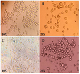
Figure 1. A) SP2/0 cells, B) Immunized mice splenic cells, C) Hybridomas on first day after fusion and D) hybridoma colonies on 15 days after fusion.
|
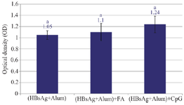
Figure 2. Optical density of all wells in three experimental groups. There were significant differences between the average OD of all wells in three groups. The highest average OD was shown in (HBsAg+Alum)+CpG group. Data is expressed as average±SD and the different lowercase letters indicates statistical significance between experimental groups (p≤0.05).
|
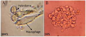
Figure 3. Optical density of positive wells in three experimental groups. There was not significant differences between the average OD of positive wells in three groups, however, the highest average OD was shown in (HBsAg+Alum)+CpG group. Data is expressed as average±SD and the same lowercase letters indicates statistical nonsignificant differences between experimental groups.
|
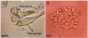
Figure 4. Single hybridoma cell (A) and single colony of specific hybridoma cell (B) after limited dilution culture.
|
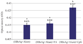
Figure 5. Average OD of all wells in three experimental groups in limited dilution culture. Average OD of all wells was significantly higher in the (HBsAg+Alum)+ CpG group than two other groups. The different lowercase letters indicates statistical significance between experimental groups (p≤0.05).
|
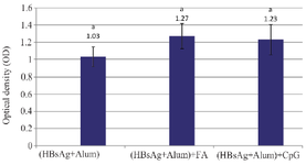
Figure 6. Average OD of positive wells in three experimental groups in limited dilution culture. There was not significant differences between averages OD of positive wells in (HBsAg+Alum) and (HBsAg+Alum)+FA and (HBsAg+Alum)+CpG groups. The same lowercase letters indicates statistical nonsignificant differences between experimental groups.
|
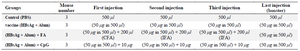
Table 1. Immunization program of mice in three groups
|
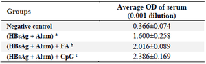
Table 2. Average OD of anti HBsAg antibody in immunized mice
Lowercase letters a, b and c letters indicates statistical differences between experimental groups.
|
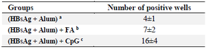
Table 3. Average number of positive wells in hybridoma culture
Lowercase letters a, b and c letters indicates statistical differences between experimental groups.
|
|