Annexin-A5 Overexpression Increases Sensitivity of MCF-7 and MCF-7/ADR Cells to Epirubicin
-
Ghasemi, Mahshad
-
Division of Medical Biotechnology, Department of Laboratory Sciences, School of Paramedical Sciences, Shiraz University of Medical Sciences, Shiraz, Iran
-
Reiazi , Niloofar
-
Division of Medical Biotechnology, Department of Laboratory Sciences, School of Paramedical Sciences, Shiraz University of Medical Sciences, Shiraz, Iran
-
Behzad-Behbahani, Abbas
-
Diagnostic Laboratory Sciences and Technology Research Center, School of Paramedical Sciences, Shiraz University of Medical Sciences, Shiraz, Iran
-
 Takhshid, Mohammad Ali
Division of Medical Biotechnology, Department of Laboratory Sciences, School of Paramedical Sciences, Shiraz, University of Medical Sciences, Shiraz, Iran, Tel: +98 0917 3121699; E-mail: takhshidma@sums.ac.ir
Takhshid, Mohammad Ali
Division of Medical Biotechnology, Department of Laboratory Sciences, School of Paramedical Sciences, Shiraz, University of Medical Sciences, Shiraz, Iran, Tel: +98 0917 3121699; E-mail: takhshidma@sums.ac.ir
-
Diagnostic Laboratory Sciences and Technology Research Center, School of Paramedical Sciences, Shiraz University of Medical Sciences, Shiraz, Iran
Abstract: Background: Multi-drug resistance is an important challenge in the chemotherapy of cancer. The role of annexin A5 (ANXA5) in the biology of cancer has been the focus of many studies. Breast Cancer (BC) is frequent cancer in women with high morbidity and mortality rate. The present study aimed to investigate the effects of ANXA5 overexpression on the anti-tumor activity of Epirubicin (EPI) in MCF-7 and MCF-7/ADR cells.
Methods: MCF-7 and MCF-7/ADR cells were transfected with the pAdenoVator-CMV-ANXA5-IRES-GFP plasmid or mock plasmid. The overexpression of ANXA5 was evaluated using qPCR. The effects of ANXA5 overexpression and EPI on the cell viability of MCF-7 and MCF-7/ADR cells were measured using an MTT assay. Cell apoptosis was measured by annexin V/7-AAD flow cytometry assay.
Results: Following the overexpression of ANXA5, the viability of MCF-7 and MCF-7/ADR was significantly decreased. Furthermore, the overexpression of ANXA5 in MCF-7 cells increased the cytotoxic effects of EPI in all doses and reduced the IC50 of EPI from 17.69 µM to 4.07 µM. Similarly, the overexpression of ANXA5 in MCF7-ADR cells reduced the IC50 of EPI from 27.3 µM to 6.69 µM. ANXA5 overexpression alone or combined with EPI treatment increased the apoptosis of MCF7 and MCF7-ADR cells.
Conclusion: The results of the present study demonstrate that ANXA5 overexpression increases the sensitivity of MCF-7 and MCF-7/ADR to EPI, suggesting a possible beneficial role of ANXA5 in the therapy of BC.
Introduction :
Breast Cancer (BC) is among the most prevalent cancer among women globally 1. Current therapies including surgical resection, chemotherapy, immunotherapy, and radiotherapy are effective in the management of patients with early and non-metastatic BC. However, Multi-Drug Resistance (MDR) and metastasis considerably reduce the efficacy of these therapeutics’ modalities 2. Therefore, many efforts have been done to understand the underlying mechanisms and to address strategies for overcoming these challenges. Several host and tumor cell characteristics are involved in MDR, most notably increased drug efflux from tumor cells that are catalyzed by MDR proteins 3. MDR proteins in BC include MDR1 protein (P-glycoprotein), Breast Cancer Resistance Protein (BCRP), and Multi drug Resistance-associated Protein-1 (MRP-1). Previous studies have revealed the contribution of MDR proteins in developing BC tumor cells to doxorubicin, paclitaxel, and etoposide 4.
Annexin A5(ANXA5) is a member of the annexin family of proteins and is well-known for its ability to bind to anionic phospholipids in a Ca2+- dependent manner 5. ANXA5 displays several activities including cell membrane repair 6, anti-coagulation 7, anti-inflammatory 6, and immunomodulatory effects 8. Cancer biology is another area of ANXA5 activity that has
been the focus of numerous researches in recent decades 9. However, contradicting data present in the literature about the function of ANXA5 in this issue. It has been reported that ANXA5 overexpression in cervical carcinoma cell lines can reduce cell proliferation and metastasis by regulating the expression of genes involved in the control of apoptosis including bax and bcl2 10. Furthermore, Wang et al showed that ANXA5 can suppress Hela cells proliferation and metastasis by regulating PI3K/Akt signal pathway 11. In a recent study, Zamani et al showed that ANXA5 overexpression can decrease migration of colorectal cancer cells through alteration in the expression of genes involved in Epithelial-to-Mesenchymal Transition (EMT) including E-cadherin, Snail, and matrix metalloproteinase -9 (MMP-9) 12. On the other hand, there is evidence that showed the stimulatory role of this protein on the viability of tumor cells. For example, it has been demonstrated that overexpression of ANXA5 promotes glioblastoma cell invasion, MMP-2 expression/activity, and chemo-resistance through a PI3K-dependent mechanism 13.
Epirubicin (EPI) is one of the anthracycline derivatives of doxorubicin which functions similarly and exhibits comparable antitumor activity compared to its parent substance, with lower heart and blood toxicity compared to doxorubicin 14. However, drug resistance is one of the major drawbacks in using EPI in the treatment of BC. It has been reported that EPI resistance is closely associated with the EMT 15. In a recent study, we showed that ANXA5 overexpression can modify the expression of genes involved in the EMT 12. To the best of our knowledge, the effects of ANXA5 on EPI resistance has not been investigated so far. To this end, we investigated the effects of ANXA5 overexpression on sensitizing MCF7 and MCF7-ADR cells, a drug-resistance cell line, to EPI.
Materials and Methods :
Cell culture: MCF7 and MCF7-ADR cell lines were purchased from the cell bank of Pasteur Institute (Tehran, Iran). The cells were kept and grown in high glucose Dulbecco's Modified Eagle's Medium (DMEM) supplemented with 10% Fetal Bovine Serum (FBS) and 1% Penicillin/Streptomycin (Gibco).
Transfection: pCMV6-AC-ANXA5-IRES-GFP plasmid harboring ANXA5 gene or mock plasmids (Origene Technologies Inc., USA) were used in this study. The recombinant plasmid was constructed in our previous study 12. MCF-7and MCF-7/ADR cells were seeded in six-well culture plates and transfected with pCMV6-AC-ANXA5-IRES-GFP plasmid harboring ANX-A5 gene or mock plasmids, using Transfectimine reagent (Dara Zistfan Eram; Shiraz, Iran) according to the manufacturer’s protocol. Briefly, a tube containing pCMV6-ANXA5 (9 µg), 200 µl of DMEM, and another tube containing Transfectimine (15 µl) and 200 µl of DMEM were prepared. Tubes were kept at room temperature for five min. Afterward, the tubes were mixed and incubated at room temperature for 20 min. The mixture was poured on the surface of the culture media, and the cells were incubated for 8 hr at 37°C in 5% CO2. At the end of this period, the medium was removed and the cells were carefully washed with Phosphate-Buffered Saline (PBS), and then cultured in fresh warm complete medium.
Real-time PCR: Real-time PCR was used to evaluate the expression of ANXA5 in treated cells and controls. Seventy-two hours after transfection, total cellular RNA was extracted using TRYzol RNA Extraction Reagent (RNXPlus, sinaclone). Complementary DNA (cDNA) was then synthesized, using PrimeScript 1st strand cDNA Synthesis Kit (Parstous, Iran), according to the manufacturer’s instructions. qPCR was performed on ABI 7500 Real Time Analyzer (Applied Biosystem, USA), using high-ROX RealQ Plus 2x Master Mix Green (Ampliqon, Denmark) and specific primers. The primers were designed by Allele ID (version 7.5) (Table 1). The experiments were performed independently three times. Pfaffl formula using β-actin as a housekeeping reference gene was applied to calculate relative changes in mRNA expression 16.
MTT assay: Seventy-two hr after transfection, the cells were treated with different doses of EPI 3.125, 6.25, 12.5, 25, and 50 µM) to compare their toxicity in MCF-7 cells with MCF-7/ADR cells. To determine possible synergistic effects between ANX-A5 overexpression and EPI, the cell lines were transfected with pCMV6-AC-ANXA5-IRES-GFP plasmid and 24 hr after transfection they were treated with different doses of EPI for 48 hr. The effects of each treatment were then determined using an MTT assay. Briefly, the cells were exposed to 5 mg/ml of Dimethyl-thiazolyl diphenyl tetrazolium bromide (MTT; Sigma Aldrich, USA) for four hours at 37°C. Afterward, 100 μl of Dimethyl Sulfoxide (DMSO; Sigma Aldrich, USA) was added to each well and the plates were left in dark for 10 min. Finally, the absorbance of each well was measured in 570 nm wavelength, using an ELISA reader instrument.
Apoptosis assay: Cell apoptosis was measured 72 hr after transfection using PE Annexin V Apoptosis Detection Kit and FACSCan flow cytometry system (BD bioscience, USA) according to the instructions provided by the manufacturer. A total number of 10,000 events were obtained for each sample within one hr.
Statistical analysis: Statistical analyses were conducted using SPSS software. Non-parametric Kruskal-Wallis was used to compare the significant difference between groups. Data are presented as mean±SD. p<0.05 was considered a significant difference.
Results :
Plasmid transfection: MCF7 and MCF7-ADR cells were transfected with pCMV6-AC-ANXA5-IRES-GFP plasmid and transfection efficiency was estimated by counting GFP-expressing cells. A high number of GFP-expressing cells was observed 24 and 48 hr after transfection, suggesting successful transfection (Figure 1). Counting the number of GFP-expressing cells in at least five fields of the cultured cells revealed that the transfection efficiency was approximately 70 and 68% for the MCF7-ADR and MCF7 cells, respectively.
Overexpression of ANXA5 in MCF7 and MCF7-ADR cells following transfection: Seventy-two hr after transfection, the expression of ANXA5 was evaluated using real-time PCR. The result showed a significant substantial increase of ANXA5 mRNA in MCF7 and MCF7-ADR cells compared to untreated control and mock cells (p=0.001) (Figure 2).
Combined effects of annexinA5 overexpression and EPI on reducing cell viability: To evaluate the possible cytotoxic effect of ANXA5 on cell viability of the MCF7 and MCF7-ADR cells, ANXA5 was overexpressed in the cells and cell viability was measured using MTT assay at 72 hr after the transfection. The results showed that ANXA5 overexpression decreased the cell viability of both cells significantly (Figure 3).
Figure 4 shows the effects of EPI on the cell viability of the MCF7 and MCF7-ADR cells in the presence and absence of ANXA5 overexpression. In the absence of ANXA5 overexpression, EPI reduced the viability of both cells in a dose-dependent manner (Figure 4). However, the sensitivity of the MCF7 cells to the cytotoxic effects of EPI was significantly higher (IC50= 17.69 μM) compared to the MCF7-ADR cells (IC50= 27.3 μM). Resistance Indexes (RI) were calculated at IC50 concentration and the data showed higher resistance of MCF7-ADR cells to cytotoxic effects of EPI in comparison to MCF7 cells (RI=1.54). To assess the effects of ANXA5 overexpression on the cytotoxic effects of EPI, ANXA5 overexpressed cells were treated with increasing concentrations of EPI and cell viability was measured using MTT assay (Figure 4). As shown in figure 4A, the overexpression of ANXA5 increased cytotoxic effects of EPI in all doses and reduced IC50 of EPI from 17.69 µM to 4.07 µM; i.e. the sensitivity of MCF cells to EPI increased about 4 folds in response to ANXA5 overexpression. Similarly, the sensitivity of MCF7-ADR cells to EPI increased about 4 folds following ANXA5 overexpression (Figure 4B), suggesting synergistic effects of EPI and ANXA5 on reducing the viability of MCF-7 and MCF7-ADR cells.
Apoptosis assay: Flow cytometry analysis using Annexin V-PE/PI method was performed to determine the role of apoptosis in the cytotoxic effects of ANXA5 and EPI. As shown in figures 5 and 6, ANXA5 overexpression and EPI at IC50 concentration caused a significant increase in the apoptosis of MCF7 (Figures 5 and 6A) and MCF7-ADR cells (Figures 5 and 6B) compared to the control cells. Combined ANXA5 overexpression and EPI treatment resulted in increased apoptosis of MCF7 (Figure 5 and 6A) and MCF7-ADR cells (Figure 5 and 6B) which was significantly higher compared to ANXA5 overexpression alone.
Discussion :
BC is known as the second most common and the first cause of cancer-related deaths among women, globally 17. The resistance to chemotherapy remains an important challenge in the therapy of patients with can cer so far. Therefore, understanding the molecular mechanisms and finding novel therapeutic methods to overcome drug resistance is of great importance. The impacts of ANXA5 on several cell signaling pathways involved in cancer drug resistance such as MAP kinase and cyclooxygenase-2 (COX-2) have been shown in previous studies 18,19. Here, we investigated the effects of ANXA5 overexpression on the sensitivity of MCF-7 and MCF-7/ADR cell lines to EPI. Based on our data, ANXA5 overexpression significantly increased the sensitivity of MCF-7 and drug resistance-MCF-7/ADR cells to EPI. Increased cell apoptosis through may involve in this process.
In the current study, we compared the effect of EPI on MCF-7 and MCF7-ADR cell viability using the MTT method. The data showed that EPI reduced the proliferation of both cell lines in a dose-dependent manner that is consistent with the results of a previous study conducted by Xiong et al 20. Comparing the IC50s of EPI (MCF7=17.69 µM vs MCF7-ADR=27.3 µM) indicated that MCF-7 cells were more sensitive than MCF-7/ADR cells to EPI. Similarly, previous studies revealed lower sensitivity of MCF-7/ADR cells to doxorubicin compared to MCF-7 cells 21,22. Wu et al reported that co-delivery of P-glycoprotein siRNA and doxorubicin can efficiently inhibit MCF-7/ADR cell viability, suggesting a role of MDR proteins in low susceptibility of MCF-7/ADR cells to cytotoxic effects of chemotherapeutic drugs 22.
To evaluate the effects of ANXA5 overexpression on cell viability, MCF-7, and MCF7-ADR cells were transfected with ANXA5 plasmid and the cell viability was measured using MTT and apoptosis assays. The results showed a significant decrease in cell viability and a marked increase in the apoptosis of both cell lines following ANXA5 overexpression. These results are consistent with the results of previous studies in cervical and prostate cell lines. In prostate cancer cell lines, auranofin, a lipophilic gold compound, decreased cell viability through the upregulation of ANXA5 expression 23. Furthermore, Li et al showed that ANXA5 overexpression inhibited the proliferation of cervical cancer cells 24. Previous studies also showed an important role of ANXA5 overexpression in meditating calcium phosphate-induced apoptosis of chondrocytes 25 and cisplatin-induced cell death of human kidney epithelial cells 26. Several possible mechanisms have been described for the apoptotic effects of ANXA5, most notably the release of apoptogenic factors such as cytochrome C from mitochondria to cytosol 19 and activation of Bak apoptotic protein through inhibition of PKCα 27. Contradictory to the abovementioned studies, cell growth-inducing effects of ANXA5 have been reported in renal cell carcinoma 28, suggesting cell-specific effects of ANXA5.
The findings of this study also showed that overexpression of ANXA5 in MCF-7 cells can reduce the IC50 of EPI from 17.69 µM to 4.07 µM which means 4 folds increased in sensitivity of MCF-7 cells to EPI in response to ANXA5 overexpression. Similarly, the sensitivity of MCF7-ADR cells to EPI significantly increased following ANXA5 overexpression. Several possible mechanisms may describe this observation. COX-2 is a regulatory enzyme that controls prostaglandins biosynthesis. COX-2 plays important role in the multidrug resistance of tumors by up-regulating the expression of MDR proteins 29. In prostate cancer cell lines, Beak et al demonstrated that ANXA5 can suppress the expression of COX-2 by downregulating of PKC-ζ-NF-κB signaling pathway. Therefore, it can be speculated that the increased sensitivity of MCF-7 and MCF7-ADR cells to EPI following overexpression of ANXA5 is attributed to COX-2 inhibition 30.
Conclusion :
In conclusion, the results of the present study demonstrated that ANXA5 can increase the chemo-sensitivity of tumor cells. These results suggest possible benefits of ANXA5 overexpression in BC therapy. Further studies using animal models are needed to confirm the results of the current in vitro study and shed light on the possible contributing mechanisms.
Acknowledgement :
This paper was a part of Mahshad Ghasemi’s MSc thesis and was supported by grant No 21378 from the Vice-Chancellor for Research Affairs of Shiraz University of Medical Sciences, Shiraz, Iran. This study was approved by the Ethic committee of Shiraz University of Medical Sciences (IR.SUMS.REC.1399. 843). The authors are grateful to all staff of the Diagnostic Laboratory Sciences and Technology Research Center and Shiraz Institute for Cancer Research Center for their technical assistance in this work.
Conflict of Interest :
The authors declare that they have no conflict of interest.
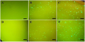
Figure 1. GFP expression was used to evaluate the efficiency of plasmid transfection. MCF7 cells showed GFP expression 24 hr (B) and 48 hr (C) after transfection. MCF7-ADR cells showed GFP overexpression 24 hr (E) and 48 hr (F) after transfection. Figure A and D illustrate the control un-transfected MCF7 and MCF7-ADR cells. Scale bar: 20 µM.
|
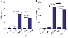
Figure 2. The expression of ANXA5 in MCF7 (A) and MCF7-ADR (B) cells compared to the control and mock group. Seventy-two hours after transfection with PCMV6-ANXA5-GFP-IRES or mock plasmids, the expression of ANXA5 was evaluated using real-time PCR in different groups and the fold change was calculated compared to that of control group. Represented data are mean ± SD of four independent experiments. Data were analyzed using the Kruskal-Wallis test and p<0.05 was considered a significant difference between the groups.
|
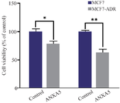
Figure 3. MCF7 and MCF7-ADR cells were transfected with PCMV6-ANXA5-GFP-IRES plasmid and their viability was evaluated using MTT assay after 72 hours. Represented data are mean±SD of four independent experiments and expressed as the percentage of cell viability in the control group. Data were analyzed using the Mann-Whitney test and p<0.05 was considered a significant difference between the groups. ANXA5 overexpression reduced cell viability in MCF7 (* p=0.01) and MCF7-ADR cells (** p=0.001) compared to that of control group.
|
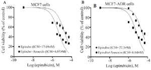
Figure 4. Effects of ANXA5 overexpression on cytotoxic effects of EPI in MCF7 (A) and MCF7-ADR (B) cells. The ANXA5 overexpressed cells were treated with various concentrations of EPI (3.125-50 µM) for 48 hr and cell viability was measured using an MTT assay. Data are mean ± SD from at least three independent experiments, each performed in triplicate, and expressed as the percentage of cell viability in control group, estimated by non-linear regression as described in methods. The IC50 of epirubicin was decreased from 17.69 µM to 4.07 following overexpression of ANXA5 in the MCF7 cells and from 27.3 to 6.64 µM to 4.07 in the MCF7-ADR cells.
|
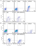
Figure 5. Annexin V-FITC/PE staining analysis of apoptosis in MCF7 and MCF7-ADR cells following transfection of AnnexinA5 and Epirubicin treatment. Analysis of apoptosis in MCF7 cells (A) and MCF7-ADR (B) transfected with AnnexinA5 or Mock plasmid vectors with or without Epirubicin treatment for 48 hr.
|
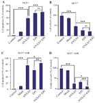
Figure 6. The effects of ANXA5 overexpression and EPI on the apoptosis of MCF7 (A) and MCF7-ADR (B) cells. The cells were transfected with the PCMV6-ANXA5-GFP-IRES and mock plasmids and treated with EPI at IC50 concentration for 48 hr. Apoptosis assay was performed using Annexin V-FITC/PE assay. Histograms showed the percentage of apoptotic cells in different groups. The presented data are mean±SD. Data were analyzed using Kruskal-Wallis’s test and p<0.05 was considered a significant difference between the groups. *p<0.05, ** p<0.01, ***p<0.001.
|

Table 1. The sequence of primers used in Real-time PCR
F: Forward Primer, R: Reverse Primer.
|
|