In Vitro Activity of Linezolid in Combination with Photodynamic Inactivation Against Staphylococcus aureus Biofilms
-
 Kashef, Nasim
Department of Microbiology, Faculty of Biology, College of Science, University of Tehran, Tehran, Iran, Tel: +98 21 61113558, E-mail: kashefn@khayam.ut.ac.ir
Kashef, Nasim
Department of Microbiology, Faculty of Biology, College of Science, University of Tehran, Tehran, Iran, Tel: +98 21 61113558, E-mail: kashefn@khayam.ut.ac.ir
-
Department of Microbiology, Faculty of Biology, College of Science, University of Tehran, Tehran, Iran
-
Akbarizare, Mahboobeh
-
Department of Microbiology, Faculty of Biology, College of Science, University of Tehran, Tehran, Iran
-
Razzaghi, Mohammad Reza
-
Laser Application in Medical Sciences Research Center, Shahid Beheshti University of Medical Sciences, Tehran, Iran
Abstract: Background: Biofilm infections are a major challenge in medical practice. Bacteria that live in a biofilm phenotype are more resistant to both antimicrobial therapy and host immune responses compared to their planktonic counterparts. So, there is need for new therapeutic strategies to combat these infections. A promising approach [known as Photodynamic Inactivation (PDI)] to kill bacteria growing as biofilms uses light in combination with a photosensitizer to induce a phototoxic reaction which produces reactive oxygen species that can destroy lipids and proteins causing cell death. PDI does not always guarantee full success, so, combination of PDI with antibiotics may give increased efficiency. This study aimed to determine if PDI was effective in the eradication of Staphylococcus aureus (S. aureus) biofilms in combination with linezolid.
Methods: The susceptibility of biofilm cultures of three S. aureus strains to Methylene Blue (MB) and Toluidine Blue O (TBO)-mediated PDI was determined alone and in combination with linezolid.
Results: Bactericidal activity (≥3 log10 reduction in viable cell count) was not achieved with MB/TBO-PDI or antibiotic treatment alone. When antibiotic treatment was combined with TBO-PDI, a greater reduction in viable count than antibiotic alone was observed for two strains.
Conclusion: This study showed that although TBO-PDI did not have good bactericidal activity against S. aureus biofilms; it increased the antimicrobial activity of linezolid against these bacteria.
Introduction :
Biofilms are microbial communities that are enclosed in a matrix of exopolysaccharide 1. This gelatinous matrix allows the growing biofilm to develop a three-dimensional structure that secures long term survival of the bacteria and makes them less susceptible to antimicrobial agents and host immune response 2. Biofilms can persist in 20 to 1000 times the concentrations of drugs that inhibit planktonic bacteria and this is due to restricted antibiotic diffusion through the matrix, slow growth rates, and induction of a resistant phenotype 3,4. Biofilms are responsible for a large number of persistent human infections such as infections of the urinary tract, middle ear, sinuses and wounds 2,5.
One promising solution to the problem posed by the reduced susceptibility of biofilms to antibiotics is Photodynamic Inactivation (PDI) 6. PDI is a process in which microorganisms are treated with a Photosensitizer (PS) and then irradiated with appropriate wavelength of visible light. The photochemical reactions generate cytotoxic Reactive Oxygen Species (ROS) which are able to exert bactericidal effect 7.
Several researches have indicated that the phototoxic effects of the PSs for eradication of biofilms are different from those of planktonic cultures 8-11. One important reason is that the penetration of the PS into the biofilms is affected by the presence of extracellular polymeric substance in the biofilms, so the phototoxic effect of the PS is reduced 9.
Some researchers have reported the effects of PDI followed by antibiotic treatment on pathogenic bacteria 12-15. According to these studies, combinations of PDI with antibiotics may give increased efficacy; so, the aim of this study was to determine whether an increased effect on eradication of Staphylococcus aureus (S. aureus) biofilms was evident if PDI was used followed by linezolid. Linezolid is an oxazolidinone antibiotic with activity against gram-positive organisms, including vancomycin-resistant enterococci, multidrug-resistant pneumococci, and methicillin-resistant S. aureus 16.
Materials and Methods :
Bacterial strains and culture conditions: The microorganisms used in this study were S. aureus UTMC 1440, S. aureus UTMC 1454 and S. aureus ATCC 25923. S. aureus UTMC 1440 and S. aureus UTMC 1454 had previously been isolated from chronic diabetic foot ulcers and characterized by conventional biochemical tests using standard methods. Bacteria were grown overnight in tryptic soy broth (Merck) under aerobic conditions at 37°C.
Assessment of biofilm formation: Assessment of biofilm formation was performed according to Sharma et al study 17.
Photosensitizer and light source: Methylene Blue (MB) (Sigma, UK) and Toluidine Blue O (TBO) (Sigma St. Louis, MO, USA) solutions were prepared fresh for each experiment in sterile PBS (pH=7.4), filter-sterilized and kept in the dark.
A 35 mW diode laser (Lasotronic-Switzerland, emitting light with a wavelength of 660 nm, fluence rate of 91 mW/cm2) and a 10 mW diode laser (MUSTANG-Russia, emitting light with a wavelength of 630 nm, fluence rate of 26 mW/cm2) was used for irradiation of samples incubated with MB and TBO, respectively.
Energy density delivered to each well was calculated using the following formula:
Energy density (J/cm2)=Power (W)×time (s)/area of each well (cm2)
The diameter of each well was 7 mm.
Susceptibility of biofilm-grown bacteria to PDI: Overnight cultures of S. aureus strains were diluted at 1:100, in TSB containing 0.25% glucose. 100 µl of the diluted bacterial suspensions and 100 µl of fresh TSB were inoculated into 96-well flat-bottomed polystyrene microplates (SPL, Korea), and incubated for 18 hr at 37°C. Following incubation, planktonic cells were removed from the microwells, 200 µl of MB/TBO at final concentration: 5, 50, and 500 µg/ml was added to each well and the plates were incubated in the dark for 10 min at room temperature. Following incubation, MB/TBO was removed from the microwells and the biofilms were washed with fresh PBS. MB/TBO-treated biofilms were irradiated with red light for 10 min (light dose: 54.6 J/cm2 and 15.6 J/cm2, respectively). Treated and untreated samples were serially diluted, plated on nutrient agar plates, and incubated for 24 hr at 37°C in the dark.
PDI experiments were also repeated with MB/TBO (final concentration: 500 µg/ml) at various incubation times (10, 15 and 20 min). All experiments were done in triplicate.
Susceptibility of biofilm-grown bacteria to PDI and linezolid in combination: Linezolid powder (Pharmacia, USA) was a gift from Dr. Alireza Foroumadi (Faculty of Pharmacy, Tehran University of Medical Sciences, Tehran, Iran).
Biofilms were allowed to form as described above and incubated with MB/TBO at a final concentration of 500 µg/ml in the dark and at room temperature for 10 min. MB/TBO-treated biofilms were irradiated with red light for 10 min (light dose: 54.6 J/cm2 and 15.6 J/cm2, respectively). 200 µl of antibiotic was added to each well at a fixed concentration of 1600 mg/L (400 times more than linezolid Minimum Inhibitory Concentration (MIC) values for these strains in planktonic forms) and plates were incubated for 24 hr at 37°C in the dark. Supernatant was removed from the microwells. Treated and untreated samples were diluted, plated on nutrient agar plates, and incubated for 24 hr at 37°C. All experiments were done in triplicate. PDI/antibiotic combinations that reduced the original inoculum by ≥3 log10 cfu/mL were considered bactericidal 18.
Statistical analysis: Values were expressed as log10 means±standard deviation. Comparisons between means of groups were analyzed using the One-Way ANOVA and Post Hoc Bonferroni tests. P<0.05 was considered statistically significant.
Results :
Biofilm formation: As indicated by the results of the crystal violet assay (Table 1), each three S. aureus strains produced biofilms [Optical Density (OD) >0.17].
Susceptibility of biofilm-grown bacteria to PDI: MB and TBO-PDI did not result in a significant reduction in viable count for any of the strains when grown in biofilms (Figure 1). Microbial reduction was not greater than 0.6 log10 with MB or 0.7 log10 with TBO. The survival of each three S. aureus strains in biofilms did not decrease with increasing PS concentrations (Figure 1) or increasing incubation time of PSs (Figure 2).
Susceptibility of biofilm-grown bacteria to PDI and linezolid in combination: Figures 3 and 4 show susceptibility of biofilm-grown bacteria to PDI and antibiotic (linezolid) in combination.
When exposed to antibiotic only, microbial reduction in comparison with the untreated control, was not greater than 0.7 log10 for each three strain. For S. aureus (ATCC 25923), the combination of MB-PDI and antibiotic resulted in a greater reduction in viable count (1.2 log10-unit reduction) than antibiotic alone (0.6 log10-unit reduction).
When antibiotic treatment was combined with TBO-PDI, a greater reduction in viable count than antibiotic alone was observed for S. aureus (ATCC 25923), and S. aureus (UTMC 1440) [2.1 and 2.6 log10- unit reduction, respectively]. Neither linezolid nor MB/TBO-PDI exhibited bactericidal activity when used alone for 3 strains. Bactericidal activity was not achieved even with PDI-linezolid combination.
Discussion :
In the present study, MB and TBO-PDI did not result in a significant reduction (≥3 log10) in viable count for any of the strains when grown in biofilms. Vilela et al also showed that microbial reduction was not greater than 1 log10 with MB or TBO 19.
Poly-N-acetyl Glucosamine (PNAG) polymer is required for bacterial adherence and biofilm formation of Staphylococcus species 20. As PNAG is a positively charged linear homoglycan, penetration of cationic PSs such as MB and TBO could be difficult through biofilms composed of PNAG. In this study, the survival of each three S. aureus strains in biofilms did not decrease even with increasing PS concentrations or increasing incubation time of PSs. So, to overcome this problem, applying other PSs may improve the efficacy of PDI on S. aureus biofilms. Other PSs such as Aminolevulinic Acid (ALA) 21, protoporphyrin-IX 22, and chlorine e6 23 have been used to evaluate the effectiveness of PDI on S. aureus biofilms. In all cases, the reduction in cell survival within biofilms and the disruption of biofilms were observed.
In this study, we demonstrated that pretreatment of S. aureus biofilms with TBO-PDI, followed by addition of linezolid at concentration significantly below the biofilm eradication concentration values, had better effect on killing of bacteria in biofilms compared to each treatment alone. In Di Poto et al study, TMP-PDI-treated S. aureus biofilms exposed to vancomycin resulted in their almost eradication. Their study showed that PDI increased susceptibility to vancomycin and it was suggested that this was as a result of the destruction of the biofilm matrix covering the bacterial cells, making them susceptible to the antibiotic 24. Cassidy et al also studied the increased bactericidal effect of PDI and antibiotic treatment in combination on Burkholderia cepacia complex strains 14. Similarly, Dastgheyb et al investigated combined use of antibiotics and meso-tetra (4-aminophenyl) porphine (TAPP) for treatment of S. aureus contamination 15.
In PDI, due to the short lifespan and limited diffusibility of singlet oxygen 25, cellular damage occurs in regions near to the PS and is not particularly targeted to structures within the bacterial cell. Therefore, the mechanism by which PDI increases linezolid susceptibility, may be due to the increased permeability of the biofilm matrix. In summary, this study showed that although TBO-PDI did not have good bactericidal activity against S. aureus biofilms, it increased the antimicrobial activity of linezolid against these bacteria.
Acknowledgement :
This work was supported by the College of Science, University of Tehran and Laser Application in Medical Sciences Research Center, Shohada Tajrish Hospital, Shahid Beheshti University of Medical Sciences.
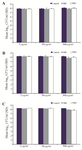
Figure 1. Effect of exposure to MB and TBO-PDI (diode laser, red light, light dose: 54.6 J/cm2 and 15.6 J/cm2, respectively) at a range of PS concentrations on killing of biofilm-grown strains. A) S. aureus (ATCC 25923), B) S. aureus (UTMC 1440), C) S. aureus (UTMC 1454).
|

Figure 2. Effect of exposure to MB and TBO-PDI (diode laser, red light, light dose: 54.6 J/cm2 and 15.6 J/cm2, respectively) at a range of incubation time of PSs (500 µg/ml) on killing of biofilm-grown strains. A) S. aureus (ATCC 25923), B) S. aureus (UTMC 1440), C) S. aureus (UTMC 1454).
|
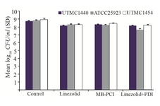
Figure 3. Effect of exposure to MB-PDI (diode laser, 660 nm, 54.6 J/cm2) and linezolid (1600 mg/L) on killing of biofilm-grown strains.
|
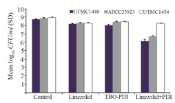
Figure 4. Effect of exposure to TBO-PDI (diode laser, 630 nm, 15.6 J/cm2) and linezolid (1600 mg/L) on killing of biofilm-grown strains.
|
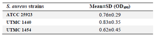
Table 1. Biofilm formation ability of two clinical S. aureus isolates and S. aureus (ATCC 25923)
|
|