The Anticancer Activity Compared Between Triptorelin and a New Gonadotropin Releasing Hormone Analogue
-
Alizadeh, Abdolali
-
Department of Organic Chemistry, School of Basic Science, Tarbiat Modares University , Tehran, Iran
-
Shamsipour, Fereshteh
-
Monoclonal Antibody Research Center, Avicenna Research Institute, ACECR , Tehran, Iran
-
 Naderi-Manesh, Hossein
Ph.D., Department of Biophysics, School of Basic Science, Tarbiat Modares University, Tehran, Iran, Tel: +98 21 88006652, Fax: +98 21 88006652, E-mail: Naderman@modares.ac.ir
Naderi-Manesh, Hossein
Ph.D., Department of Biophysics, School of Basic Science, Tarbiat Modares University, Tehran, Iran, Tel: +98 21 88006652, Fax: +98 21 88006652, E-mail: Naderman@modares.ac.ir
-
Department of Biophysics, School of Basic Science, Tarbiat Modares University , Tehran, Iran
Abstract: Gonadotropin releasing hormone (GnRH) plays a key role in reproduction. This decapeptide is synthesized and released by hypothalamus and induces the pituitary gonadotrop cells to release pituitary gonadotropin hormones. In some extrapituitary compartments GnRH and its receptor act as part of the autocrine regulatory system of cell proliferation. The anticancer activity of GnRH and its analogues has been observed by many researchers. In this study the anticancer activity of a new analogue of GnRH and triptorelin was investigated by cell proliferation assay. Results indicate that proliferation of human breast and ovarian cancer cell lines are dose-dependently inhibited. The inhibitory efficiency of the new analogue is proved to be higher than the original triptorelin. In addition to its antimitogenic activity, evidence was found for the involvement of the apoptotic mechanism in the action of the new analogue and triptorelin. In conclusion, the new analogue can be considered as a good pharmaceutical candidate.
Introduction :
Gonadotropin releasing hormone (GnRH) is the central regulator of the reproductive hormonal cascade. This decapeptide is syn-thesized and released by hypothalamic secretory neuron which is delivered to the pituitary gland via hypophyseal portal blood system. Interaction of GnRH with its receptors on the pituitary gonadotrop cells induces the release of pituitary gonadotropin hormones, which in turn regulate gonadal steroidogenesis and gametogenesis in both sexes (1). Therefore, GnRH analogues have been used in the assisted reproduction (in vitro fertilization and embryo transfer), treatment of infertility due to polycystic ovarian diseases and fibroids. Also the effi-ciency of analogues of GnRH for the treat-ment of children with precocious puberty, endometrial carcinoma, estrogen-dependent breast cancer and prostate cancer is well established (2). There is growing evidence of autocrine/ paracrine GnRH systems in human reproductive tissues (3-6). Recent studies sug-gest that about 50% of breast cancer and 80% of ovarian cancer cell lines express high-affinity binding site for GnRH and its analogues as part of the autocrine regulatory system of cell proliferation. Therefore anti-cancer activity of GnRH analogues has been observed by many others (7-11).
Since 1972 systematic work has been preceding to synthesize agonistic and antag-onistic analogues of GnRH. A powerful interest in medical applications of GnRH derivatives stimulated this undertaking. Thus, the intense activity that has occurred in this field was caused by the desire to synthesize super active analogue with prolonged biologic activity. Many agonistic and antagonistic ana-logues more potent than the parent hormone have been made. Several of these analogues such as triptorelin, goserelin, leuprorelin and buserelin are being used clinically and the list of their applications is steadily expanding (2).
Considerable effort has been devoted to the synthesis of peptidomimetic structures to overcome the unfavorable properties and a therapeutic deficiency of peptides (12,13). Among them is insertion of some chemical groups in the peptide sequences to increase their activity efficiency. In the present work the anticancer activity of a new GnRH ana-logue was investigated in comparison with triptorelin. The new analogue structure is similar to triptorelin with two extra chemical groups inserted between Leu7 and Arg8 in order to increase peptide hydrophobic prop-erties (Figure 1). Based on previous report on the molecular mechanism of ligand inter-action with the GnRH receptor (14), the hydrogen bonding and p-stacking interaction play main roles in GnRH and its receptor interaction. Therefore we expect stronger interaction between new analogue and GnRH receptor leading to greater peptide activity.
Materials and Methods :
Material
[D-Trp6] LHRH (Triptorelin) was synthe-sized in our laboratory by solid phase method and the new analogue was kindly provided by Dr Balalaie et al (15). Human breast cancer cell line (T47D) and ovarian cancer cell line (OVCAR3) were obtained from Pasteur Insti-tute of Iran. RPMI 1640 and Fetal Bovine Serum (FBS) were purchased from Gibco (USA) and Biosera (UK). Multiple 6 well-dishes were Greiner bio-one (Germany) prod-uct. Annexin V-FITC apoptosis detection kit was obtained from BD Bioscience, Pharmin-gen (US).
Cell culture
Human breast cancer cell line (T47D) and ovarian cancer cell line (OVCAR3) were maintained in RPMI 1640 medium supple-mented with 10% heat-inactivated bovine serum albumin and kept at 37 °C in a humidi-fied 5% CO2 atmosphere.
Cell proliferation assay
To determine the anti-proliferation activity of the new analogue, the dose-dependent proliferation experiments were performed. In brief following steps were carried out: 2×10⁴ cells of each cell line were plated in multiple 6 well-dishes. After 24 hr, the cells were treated with 10-11, 10-9, 10-7and 10-5 M con-centrations of each peptide. This treatment was repeated three times during six days. On the sixth day, cells were trypsinized and after Trypan-blue staining, the viable cells were counted with Neubauer-type hemocytometer and the data was expressed as the percentage of control. All proliferation assays were performed at least three times.
Apoptotic assay
Cytomorphological changes were observed only in treated OVCAR3 cells in 10-9 M concentration of triptorelin with an Olympus phase-contrast microscope. Cell death by apoptosis was confirmed using Annexin V-FITC apoptosis detection kit with a vital dye such as Propidium- Iodide (PI) according to the manufacturer?s instructions. Briefly, OVCAR3 adherent cells that were treated with triptorelin and the new analogue were washed with the culture medium. Surface exposure of Phosphatidyl Serine (PS), as a plasma membrane asymmetry in apoptotic cells, was detected by adding annexin V-FITC to the culture medium in a final concentration of 5 µg/ml. The cells incubated for 5 min at room temperature, then PI was added (1 µg/ml) (16,17). After rinsing with culture medium to remove excess dyes, the cells were observed by fluorescence microscope (Olympus).
Statistical analysis
The data from three or four dose-dependent experiments were tested by ANOVA followed by post-hoc analysis (Duncan test). All analy-sis performed with SPSS software.
Result :
In T47D and OVCAR3 cell lines, prolifera-tion was dose-dependently inhibited with different concentrations (10-11, 10-9, 10-7 and 10-5 M) of the new analogue and triptorelin (Figures 2 and 3). The anticancer activity of the new analogue was higher than triptorelin at all concentrations. Table 1 gives a compari-son of the cell number percent of cancer cell lines after treatment with triptorelin and the new analogue.
After treatment of OVCAR3 cell line with the new analogue and triptorelin, some mor-phologic signs of programmed cell death could be detected by invert phase contrast microscope. The vacuoles in the cytoplasm, rounding-up of cells, apoptotic bodies and bleb formation were seen as characteristic signs of the apoptotic process, while control cells had normal morphology (Figure 4). Apoptosis was also confirmed by annexin V-FITC apoptosis detection kit. OVCAR3 cells with annexinV-/PI- were alive, annexin V+/PI- were undergoing early apoptosis and annexinV+/PI + were either in the end of apoptosis or were dead (Figure 5).
Discussion :
The anticancer activity of the new analogue was higher than triptorelin at different concentrations. The improved activity of the new analogue is probably due to its stronger interaction with the GnRH receptor. Söderhäll et al reported that the insertion of hydropho-bic groups in GnRH sequence would increase the p-stacking interaction of the peptide with the hydrophobic pocket of GnRH receptor and therefore improved its activity (18). Further-more the presence of such chemical groups in the new analogue sequence is thought to increase the protease stability of the new analogue and therefore increase its biological activity.
The results of our proliferation assay correspond to similar observations obtained by other groups and are explained by the fact that GnRH and its receptor are parts of the negative autocrine regulatory system of cell proliferation (7-10). The most important features of GnRH signaling in tumors are the inhibi-tory interference with mitogenic pathway that results in antiproliferative actions. This peptide activates a protein tyrosine phospha-tase that could inhibit the mitogenic signal transduction of growth factor receptors and therefore down regulates the cell proliferation (10). In another way our findings support previous studies that some GnRH analogues can prompt apoptosis in breast, ovarian, endometrium and prostate cancer cell lines (1,19-28). However the mechanism underlying the apoptotic effect of the analogues in the human counterpart is not fully known.
In conclusion our results show that the anticancer activity of the new analogue is more than triptorelin and it seems to be due to the known mechanisms of GnRH effect on extrapituitary compartments that was ex-plained before. Therefore this new analogue can be considered as a good pharmaceutical candidate.
Acknowledgement :
We appreciate the helpful discussion and comments given by Dr. Sadjady. The authors also express their gratitude to the Research Council of Tarbiat Modares University.
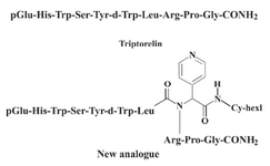
Figure 1. Chemical structures of the triptorelin and the new analogue
|
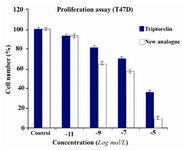
Figure 2. Effect of 6 days of treatment with different concentrations of triptorelin and the new analogue on T47D cell line. Each column represents the cell number percent in comparison with the control. The data are representative of three independent experiments. Analysis of variance: P<0.01
|
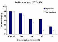
Figure 3. Effect of 6 days of treatment with different concentrations of triptorelin and the new analogue on OVCAR3 cell line. Each column represents the cell number percent in comparison with control. The data are representative of three independent experiments. Analysis of variance: P< 0.01
|
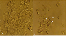
Figure 4. (a) Normal OVCAR3 cell line. (b)Apoptotic forms of OVCAR3 cell line observed by invert phase-contrast microscope. Arrows show the morphologic signs of programmed cell death in early apoptosis
|
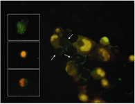
Figure 5. Apoptotic forms of OVCAR3 cell line observed by fluorescence microscope. OVCAR3 cell line was stained with annexinV-FITC/PI. Arrows show the plasma membrane asymmetry in early apoptotic cells. Boxes show different stage of apoptosis such as early and late / already dead cells
|
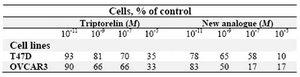
Table 1. Cell number percent of cancer cell lines after treatment with (10-11, 10-9, 10-7, and 10-5 M) concentration of triptorelin and the new analogue
|
|