Activity of Citrus aurantium and Lavandula angustifolia in Alzheimer’s Disease Symptoms in Male Wistar Rats
-
 Arasteh, Amir
Department of Biology, Rasht Branch, Islamic Azad University, Rasht, Iran, Department of Biology, Rasht Branch, Islamic AzadTel: +98 13 33423308; E-mail: arasteh@iaurasht.ac.ir
Arasteh, Amir
Department of Biology, Rasht Branch, Islamic Azad University, Rasht, Iran, Department of Biology, Rasht Branch, Islamic AzadTel: +98 13 33423308; E-mail: arasteh@iaurasht.ac.ir
-
Karimpour, Morteza
-
Department of Biology, Rasht branch, Islamic Azad University, Rasht, Iran
-
Fallah, Faezeh
-
Department of Biology, Islamic Azad University, Central Tehran branch, Tehran, Iran
-
Kiani, Sara
-
Department of Biology, Islamic Azad University, Central Tehran branch, Tehran, Iran
-
Kakavan, Maedeh
-
Department of Medical Microbiology, Faculty of Medicine, Mazandaran University of Medical Sciences, Sari, Iran
Abstract: Background: Alzheimer's Disease (AD) is one of the most prevalent chronic neurodegenerative disorders. The present study aims to better understand the mechanism by which Citrus aurantium (C. aurantium) and Lavandula angustifolia (L. angustifolia) hydro–alcoholic extracts were used to treat AD and anti–oxidant issues in a laboratory model.
Methods: 15 male Wistar rats, weighing 250±20 gr, aged 6–8 weeks, were used. Amyloids in the brain were found and identified using the shuttle box and Congo red test. ELISA testing for norepinephrine and serotonin, Superoxide Dismutase (SOD), Malondialdehyde (MDA), and Real–time PCR for expression of the APP and GLT1 genes were done.
Results: The shuttle box test demonstrated that AD produces behavioral harm, since it significantly reduces passive avoidance learning. The Congo red test revealed that the AD models had much more amyloid beta in their brain tissue than the control. Norepinephrine levels were also decreased by using both extracts in test group. Treatment with both extracts led to a substantial rise in SOD activity and fall in MDA concentration.
Conclusion: The gene expression data indicated that the relative expression of the APP and GLT1 genes was shown to be lower in the groups treated with both extracts. C. aurantium and L. angustifolia may therefore offer a multi–target treatment strategy for AD, which calls for more research in this area.
Introduction :
One of the most prevalent chronic neurodegenerative disorders, Alzheimer's Disease (AD), is characterized by neural plaques and neurofibrillary tangles as pathological signs. Both the hyper–phosphorylation of tau protein linked with microtubules in neurons and the accumulation of amyloid beta peptide (Aβ) in brain tissue are responsible for these symptoms. The amyloidogenic processing is influenced by a variety of genetic and environmental variables 1. Aβ (abeta) accumulation in the brain starts a pathogenic cascade that eventually results in synaptic dysfunction and loss, neuronal death, and finally cognitive failure. Inflammation and oxidative stress are also a part of this process, which contribute to neurological dysfunction 2-4. The only available treatments for AD are one partial N-Methyl-D-aspartate (NMDA) receptor antagonist (memantine) and four cholinesterase inhibitors (tacrine, rivastigmine, donepezil, and galantamine).
Herbal and medicinal extracts are also being studied in addition to therapeutic substances. These studies, which include the incidence and prevalence of AD in many cultures and nations, are based in part on nutritional and epidemiological investigations. For instance, a considerable reduced incidence of AD (4.4-fold) was seen among older Indians compared to an American reference sample in a major epidemiologic investigation 5.
In the primary healthcare system, medicinal plants are seen as being quite significant. Numerous herbal therapies have been examined in clinical studies and other AD-related models, and they have all demonstrated positive results 6.
Citrus aurantium var. amara L. (C. aurantium) (www.theplantlist.org, record 28100679), the plant known by its scientific name, is native to Asia's tropical regions; however, it may also be found in other tropical and subtropical areas. In Asia, C. aurantium is used as a brew or tea and C. aurantium sweat. The sweat of C. aurantium is used in salad dressing, as an air freshener and in the bedroom for better sleep. In Iran, to reduce the complications of AD in the elderly, plant syrup containing one third of a glass of C. aurantium sweat, one third of water and one third of rose water is used. Blossom of the orange tree, is widely used in perfumery and making essential oils, all kinds of seasonal drinks, and preparing jam. This herb is widely utilized in the treatment of several ailments and is freely accessible 7. Flavonoids, including hesperidin, naringin, and alkaloids, primarily synephrine, are the primary physiologically active components of C. aurantium and have positive medicinal benefits on human health. These substances have the reputation of a novel drug due to a variety of therapeutic benefits including disinfectant, anticancer, anti–inflammatory, and anti–oxidant, as well as its involvement in the treatment of cardiovascular, mental, and neurological problems 8.
Another plant whose anti-Alzheimer effects were investigated in this research is Lavandula angustifolia (L. angustifolia) Mill (www.theplantlist. org, record 108971). This plant (English name: Lavender), is widely used in Iran and Mediterranean regions to reduce Alzheimer's complications. In Iran, L. angustifolia brew is used to prevent and reduce the side effects of AD. It has been demonstrated in multiple researches 9 to have a variety of therapeutic benefits, including sedative, anticonvulsant, analgesic, and local anesthetic properties. Additionally, the nervous system is soothed and relaxed by this herb 10. In Alzheimer's patients, the amount of norepinephrine and Malondialdehyde (MDA) (an indicator of oxidative stress) increases, but the concentration of serotonin and the activity of the Superoxide Dismutase (SOD) enzyme decrease. Also, the expression of the Glutamate transporter-1 (GLT1) gene increases, and promising results were obtained in all cases with the use of medicinal plants. The present research aims to better understand the mechanism of action of C. aurantium and L. angustifolia hydro–alcoholic extracts in treating anti–oxidant issues typical of AD in an induced laboratory rat model. This understanding may protect the biodiversity rights of indigenous people and open the door to potential therapeutic applications.
Materials and Methods :
Preparation of herbal extracts: Identified and documented genus and species of C. aurantium and L. angustifolia plants were purchased from Medicinal Plants Research Center, Tehran University of Medical Sciences. The extraction was performed by the maceration method (ethanol 96%). for both plants. For example, 10 g of the dried plant powder was poured into 200 ml of 96% ethanol solution. The solution was stirred for 24 hr and filtered through a filter paper to obtain the extract.
GC–MS analysis: GC/MS analyses were performed by 7890B series gas chromatography device (Agilent Company of America), Column: 30 mm×0.25 mm and 0.25 mm film thickness. For L. angustifolia, the oven temperature program started from 60-220°C for 20 min at a rate of 6°C/min. Transfer line temperature was 280°C; injector temperature was 250°C. Components were detected by comparing the resulting spectrum to the mass pattern found in the analyzer library 11. For C. aurantium, an aliquot of 10 µl was placed in GC-MS syringe and oven temperature of 60°C for 10 min. The temperature was increased to a final temperature of 250°C in rate of 5°C/min. Injector and detector temperatures were 230°C and 300°C, respectively 12.
Animal studies, model development and treatment: 15 male Wistar rats, weighing 250±20 g, were acquired from the Pasteur Institute of Iran for this investigation. They were housed at a temperature of 23±3°C and a humidity of 50±10 throughout the experiment. Up to the completion of the treatment, the rats had unrestricted access to animal food, including water and special rat chow. The rats' sleeping and waking times were fixed to be 12 hr in a dark environment and 12 hr in light, and were categorized as shown in table 1. The research was conducted in accordance with accepted principles for laboratory animal use and care.
Rats were put to sleep using ketamine (100 mg/kg) and xylazine (10 mg/kg) before being moved to stereotaxic equipment to produce the model (RWD, 0755-86111281). The Alzheimer's model was injected and generated using β-Amyloid Peptide (1-42) (Sigma, CAS number: 107761) by injecting in the CA1 region of the hippocampus with the coordinates utilized from the Paxinos atlas (ML=-2.6, AP=-3.8, DV=2.2) and a concentration of 5 µg/μl and 2 μl on each side of the hippocampus using a 5 μl syringe (Hamilton, USA) 13. Following the model's introduction, 200 μl of C. aurantium and L. angustifolia extracts with concentration of 100 mg/kg were administered by gavage over 4 weeks, five days per week 14-17. The behavioral shuttle box test was conducted 30 days following the last treatment. The rats were subsequently put to death with carbon dioxide gas, and the brain tissue was removed and fixed in 10% formalin 13.
Shuttle box test: A shuttle box device (type ST–500) with two chambers-one light and one dark-and bottoms wrapped in steel wire with a diameter of 1 to 2 mm was used for this test. Also, in the dark chamber, a light shock of 75 mA and alternating current for three s was delivered to the mouse's leg just once using an electric current generator. The mice were all first placed in this room for ten min to become used to the open guillotine door, after which they were all moved to the light chamber, and as soon as they entered the dark chamber, the guillotine door was shut and the mice's legs were shocked with electricity. Mice were tested for passive avoidance memory 24 hr later, and the delay time for entering the dark compartment was assessed in seconds 18.
Congo red test: Once the tissue had been verified, ethanol was employed to dehydrate the subject (starting from low degrees to absolute alcohol). The alcohol was then eliminated by xylol. The samples were then mounted on a silanized slide after being microtome-cut to a thickness of 5 πm. The dehydration procedure and deparaffinization of the slides were done first. They were then immersed in Congo Red 1 solution (Sigma-C-6277) for 30 min, rinsed with distilled water, and then immersed 5-10 times in an alkaline alcohol solution (1% sodium hydroxide and 50% alcohol, Sigma-S0899). After washing the slides in water for 5 min, hematoxylin (Sigma-H9627) for 30 s, and ammonium hydroxide (Sigma-1336-21-6) for 30 s were used and they were once again put in water for washing. Dewatering and clarifying were completed in the end, and an optical microscope (LABOMED) was used to capture images 19.
ELISA test for norepinephrine and serotonin: One ml of Ripa buffer (Cat. NB: DB9719) was used to homogenize about 100 mg of tissue before centrifuging it for protein extraction. The BCA test was used to identify the supernatant solution that contained protein. After creating the standard graph, the Biotek-refelx800 (US) ELISA equipment was used to evaluate the amounts of norepinephrine and serotonin protein in the samples according to the ELISA kit's methodology 20.
Superoxide dismutase (SOD) and malondialdehyde (MDA) assay: 100 mg of tissue was combined with 1 ml of PBS (Cat. NB: A-0018) before being homogenized and centrifuged. Using the SOD kit (CAT NO: ZB-SOD), the SOD test was conducted on the supernatant of the samples following the kit's instructions. Using a tetrazolium salt that, when reduced with superoxide anion, yields a water-soluble formazan dye, this kit assessed SOD activity. The quantity of produced color was quantified at a wavelength of 440-460 nm with a Biotek-refelx800 (US) ELISA equipment when it was discovered that the presence of SOD in the media inhibits the rate of formazan production. The material was extracted using Ripa buffer, and the MDA kit (ZB-MDA) was used under the kit's instructions to test MDA. Then, using a Biotek-refelx800 (US) ELISA equipment, the quantity of color generated was measured at a wavelength of 535 nm 21.
Expression of Amyloid precursor protein (APP) and Glutamate transporter-1 (GLT1) genes: Each sample's total RNA was manually extracted using Triazol (Qiazol, Kiazist) following the manufacturer's instructions from 50 mg of tissue. Simply, 500 ng of RNA from each sample was added to the cDNA synthesis procedure using the Easy cDNA Synthesis Kit (Parstous). Afterwards the extracted RNA was read at a wavelength of 260 nm and a ratio of 260/280 and 230/260 was checked to ensure its quality and purity. Primer3 software was used to create primers for genes (Table 2). Then, using a real-time PCR thermocycler (ABI Stepone, US) and SYBR Master with high Rox (2X) (Addbio), relative changes in the expression of the APP and GLT1 genes in various groups as compared to the control group and GAPDH internal control gene were evaluated 22.
Software called Graph Pad Prism version 9 was utilized to evaluate the tests' statistical significance. ANOVA and Tukay's multiple comparison test were employed in one way. A p-value of 0.05 and a 95% confidence interval were taken into account. Each group included three tested samples. Three repetitions for each sample were run through each test. The results were expressed as mean ± standard error (mean ± SE).
Results :
Phytochemical evaluation: The results of GC–MS for C. Aurantium and of L. angustifolia are shown in tables 3 and 4. As shown in the table 3, D-Limonen was reported as the active constituent of the C. Aurantium hydro-alcoholic extract. Also, the eucalyptol was reported as the active constituent of the L. angustifolia.
Shuttle box behavioral test: The findings demonstrated that model induces behavioral impairment so that passive avoidance learning is significantly less than in the control group. Figure 1A, shows that on the first day of measurement, the model group's initial delay time to enter the dark room after the induction of an electric shock was shorter than that of the control group, which is statistically significant at p<0.05. The decrease in the treated groups when compared to the control is not statistically significant, but when compared to the model group, particularly with the treatment using both extracts, it resulted in an increase in learning, which is statistically significant. The experiment was measured again on the second day, and Figure 1B, depicted the same procedure for all control and treatment groups with additional seconds.
To evaluate memory recovery, the Time of Dark Chamber (TDC) was also investigated. Figure 2 shows a statistically significant (p<0.05) increase in TDC in the model group when compared to the control groups. In comparison to the Alzheimer's group, treatment with C. aurantium and L. angustifolia and both medications simultaneously decreased the amount of time spent in the dark room. This difference is statistically significant at p<0.05 for the L. angustifolia group and both extracts.
Congo red test: Amyloids in brain tissue were found and identified using the Congo red staining method. The outcomes of brain tissue staining in various groups have been shown in figure 3. Figure 4 compares the groups based on the three times each sample's amyloid plaque count was counted. The graphic shows that the Alzheimer's model mice had more amyloid in their brain tissue than the control and other treatment groups. The maximum significance is p<0.001 compared to each extract with the model group in the groups treated with C. aurantium and L. angustifolia extracts in terms of lowering the amount of amyloid compared to the model group. There are no significant variations between the two extracts.
ELISA test for norepinephrine and serotonin: As shown in figure 5A, after illness induction, there is a significant increase in norepinephrine protein levels in the model group compared to the control group (p<0.001). In the groups treated with C. aurantium and the two extracts, there was a reduction in this protein when compared to the model group; this reduction in protein is significant when compared to the model group. The differences between the therapy groups are negligible. Additionally, figure 5B, shows that following illness induction, the model group's level of serotonin protein decreased significantly (p<0.001) compared to the control group. There is hardly any distinction across the treatment groups. Furthermore, figure 5B demonstrates that, in comparison to the control group, the model group's level of serotonin protein reduced dramatically (p<0.001) after illness induction.
Superoxide dismutase (SOD) and malondialdehyde (MDA) tests: As shown in figure 6A, when compared to the control group, the model group's SOD activity was significantly lower (p<0.001). When compared to the model group, treatment with L. angustifolia extract and both extracts combined significantly increased SOD activity. Among the groups treated with one extract, the increase in SOD activity in the group treated with both extracts relative to the model group showed maximum significance. As shown in figure 6B, in comparison to the control group, the MDA concentration in the model group has significantly increased (p<0.001). MDA concentration was significantly lower after treatment with each extract separately and in combination than it was in the model group. Among the groups treated with one extract, the reduction in MDA levels in the group treated with both extracts relative to the model group showed maximum significance.
Expression measurement of APP and GLT1 genes by Real-time PCR method: As shown in figure 7A, in comparison to the control group, there is a significant increase (p<0.01) in the relative expression of the APP gene in the model group. In comparison to the model group, the groups treated with individual and combined extracts showed a drop in the relative expression of this gene. In the C. aurantium group and both extracts together, this decrease is significant. Figure 7B displays the level of GLT1 gene expression in different groups. In comparison to the control group, the relative expression of this gene in the model group has dramatically risen (p<0.01). The relative expression of this gene relative to the model group has significantly decreased in the L. angustifolia group and both extracts combined after the effects of the extracts both alone and together. The relative level of expression of both genes has been impacted by both extracts.
Discussion :
Numerous studies have demonstrated that food and nutrition can positively influence various pathophysiological consequences in AD so far. Additionally, some studies have suggested that dietary components such as polyphenols may be useful in avoiding and postponing age-related diseases 23,24. C. aurantium is a traditional herbal remedy that is frequently used as both an anti-oxidant and an anticancer agent 25. Additionally, L. angustifolia is recommended in conventional medicine as a therapy for AD. The therapeutic medications employed are mostly anti-oxidants, anti-inflammatory drugs, cholinergic agonists, estrogen, neurotrophic factor, etc. due to the numerous pathogenic factors in AD 26. Although organic synthesis can yield two or more pharmacophores related to multifunctional AD inhibitors, the implementation of this strategy is greatly hampered by unpredictable side effects. As a result, researchers' attention has gradually shifted away from organic synthesis and toward multifunctional compounds in natural products 27.
As the findings of the current study demonstrated, the behavioral test revealed that although the decrease in the treated groups when compared to the control is not statistically significant, the treatment with both extracts specifically resulted in an increase in learning that is statistically significant when compared to the model group. As the behavioral studies by Zhang et al indicated, Rhodiola crenulata Extract (RCE) can enhance cognitive abilities in Aβ1–42 induced AD mice models, including learning and memory 28. According to Rabies’s study the shuttle box results showed that the initial delay in the Alzheimer’s mouse model that was treated with Cyperus rotundus ethanolic extract was significantly decreased 29. The Aβ plaque system plays a role in neuropsychiatric processes including memory and learning. Disorders of learning and memory are usually caused by an increase in Aβ in certain brain areas 30. The findings of this research indicated that a significant increase exists in the level of Aβ in the mouse models' brains, which might be the reason for the cognitive deterioration found in the current study. These defects were corrected by treatment with C. aurantium and L. angustifolia extract, indicating that lavender and orange spring may improve the Aβ plaque system's functioning. As a result, C. aurantium and L. angustifolia may enhance learning and memory in behavioral tests through a variety of processes. The findings of Congo red staining of the brain tissue indicated that the groups treated with C. aurantium and L. angustifolia extracts had the maximum significance at p<0.001 compared to each extract alone with the model group in terms of lowering the quantity of amyloid plaques. Similarly, Dhanasekaran et al demonstrated using Congo red staining that a dosage of 5.0 mg/kg of Centella asiatica and a long-term treatment plan can diminish fibrillar amyloid plaques 19. Additionally, research by Soheili's team showed that L. angustifolia might significantly decrease the number of amyloid plaques in mice used as an Alzheimer's model 31.
Numerous practical research has shown that deficiencies in the anti-oxidant defense system and oxidative stress associated with Aβ are important factors in the pathophysiology and etiology of AD 9. Compared to the model group, the findings of the current study indicated that treatment with L. angustifolia extract and both extracts combined significantly increased SOD activity. Among the groups treated with one extract, the increase in SOD activity in the group treated with both extracts relative to the model group showed maximum significance. Additionally, compared to the model group, treatment with both individual and combined extracts resulted in a significant drop in MDA levels. Zhang et al demonstrated that RCE therapy might stop MDA accumulation and the reduction in SOD activity in AD mice models. The present findings are also in line with earlier research, which found that in a rat model of streptozotocin-induced hippocampus cell injury, MDA levels considerably rose along with significant deficits in learning and spatial memory, and the pretreatment with Rhodiola rosea extract greatly reduced these aberrations 32.
The findings of the ELISA test revealed that a significant increase exists in the protein level of norepinephrine in the model group compared to the control after the illness was induced. This protein was lower after treatment with each extract alone and after treatment with both extracts compared to the model group. Additionally, after illness induction, the model group's level of serotonin protein decreased significantly (p<0.001) compared to the control group. This protein was significantly elevated following treatment with each extract alone as well as treatment with combined extracts in comparison to the model group. In the study conducted by Rapaka et al, pathological and behavioral changes caused by AL-induced Alzheimer's mice were associated with a decrease in dopamine and serotonin levels in the cerebral cortex and hippocampus, and treatment with Benincasa hispida hindered this in the AD model 33. Elsawi's study also demonstrated that Lagerstroemia indica extract might change the levels of the hormones norepinephrine and serotonin in the Alzheimer's model group in comparison to the control group, respectively 34.
In comparison to the model group, the findings reveal that the relative expression of the APP gene in the groups treated with the extracts individually and together demonstrate lower expression levels. In the C. aurantium group and when the two extracts are combined, this decrease is significant. Furthermore, the L. angustifolia group and both extracts taken combined showed a significantly lower expression of GLT1 following the effects of the extracts than the model group.
Similarly, Yaghmaei group research showed that silymarin extract treatment also lowers the number of amyloid plaques in the brain. Moreover, silymarin was able to reduce APP expression as seen by the comparison of treated and untreated groups' APP gene expression 35. Ji et al also discovered that Akt phosphorylation was necessary for insulin stimulation to enhance total and surface GLT1 expression in cultured astrocytes, which was linked with KBBP expression and GLT1 mRNA transcription 36. According to abundant evidence, oxidative stress-induced abnormal and excessive Ca2+ release from the Endoplasmic Reticulum (ER) via the Ryanodine Receptor (RYR) which then plays a significant role in memory loss and cognitive dysfunction in AD patients through activation of the enzyme calcineurin phosphatase and increased activities of the enzymes β-calcineurin and γ-secretases in the generation and deposition of Aβ42 senile plaques and the APP gene. Inhibiting excessive Ca2+ release from the ER, as well as RYR over-activation and calcineurin activation in hippocampus neurons, may therefore be viable therapeutic targets for the treatment of AD 37.
Conclusion :
The aim of the present study was to better understand the mechanism of action of two plants, C. aurantium and L. angustifolia, as widely used ethnic herbal medicines, which are widely used in Iran and Mediterranean regions 38 to reduce the complications of AD. The results of this research showed that the C. aurantium and L. angustifolia are able to modulate the expression of APP and GLT1 proteins, decrease oxidative stress, protect neurons from degeneration or apoptosis, and greatly enhance the cognitive performance of AD rat in the behavioral test. Therefore, C. aurantium and L. angustifolia provide a multi-target therapeutic strategy for AD and can be introduced as nootropic agents to reduce the complications of cognitive diseases such as AD.
Acknowledgement :
The authors of this article would like to express their deepest gratitude to Mr. Talebi, who helped to improve research quality, as well as Histogenotec Research Laboratory. The costs are borne by the authors.
Conflict of Interest :
The authors declare that they have no conflict of interest.
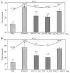
Figure 1. Initial delay time to enter the dark room after the induction of an electric shock. A) the amount of step through latency (STLa) in seconds on the first day, B) the amount of step through latency (STLr) in seconds on the second day.
|
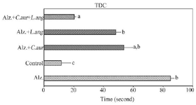
Figure 2. Time of Dark Chamber (TDC) in seconds.
|
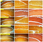
Figure 3. The Congo red staining images of a cross-section of brain tissue in mice of different groups, as shown in the figure, with the use of C. aurantium and L. angustifolia extracts. the number of amyloid plaques (denoted by white arrows) has decreased significantly compared to the sample with Alzheimer's disease. This reduction has been more with the simultaneous use of two extracts. Scale bars for the first column from the left are 200 µm and for the second and third columns are 100 µm.
|
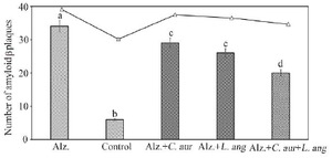
Figure 4. The number of amyloid plaques in different groups, similar signs are not significantly different. Non-similar signs have a statistically significant difference of p<0.05.
|
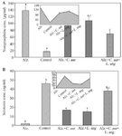
Figure 5. Norepinephrine (A) and Serotonin (B) protein expression levels in different groups, similar signs are not significantly different. Non-similar signs have statistically significant differences (p≤0.001).
|
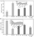
Figure 6. SOD activity (a), and MDA concentration (b), in the studied groups, Similar signs are not significantly different. Non-similar signs have statistically significant differences (p≤05).
|
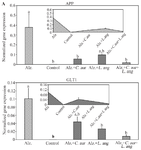
Figure 7. Changes in the relative expression of (A) APP and (B) GLT1 genes in different groups, Similar signs are not significantly different. Non–similar signs have statistically significant differences (p≤0.005).
|
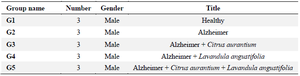
Table 1. The studied groupings for investigation of C. aurantium and L. angustifolia
|
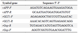
Table 2. Primer sequence designed by primer 3 software
|
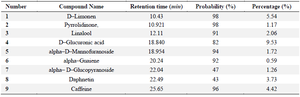
Table 3. Chemical composition of Hydro–alcoholic extract of C. Aurantium
|
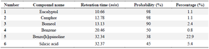
Table 4. Chemical composition of Hydro–alcoholic extract of L. angustifolia
|
|